ECGbook.com

Making Medical Education Free for All

ECGbook.com

Making Medical Education Free for All

Home /

First Degree AV block

First degree AV (heart) block

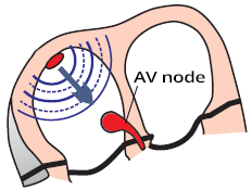
Atrioventricular (AV) Node

Atrioventricular (AV) node conduction
  • In sinus rhythm, impulses are generated regularly (about 60/min.) in the SA node
    • Each impulse spreads through the atria (P wave) to the AV node
  • In the AV node, the impulse slows down by about 0.1s
    • During this time, the atria pump blood into the ventricles
    • Then the impulse continues to the ventricles (QRS complex)

PQ Interval

AV node conduction, PR interval, PR segment
  1. Impulse originates in SA node
    • When it moves to the atrial myocardium, it starts generating the P wave
    • Simultaneously, it spreads through the conduction system towards the AV node
      • The impulse in the conduction system does not create a curve
  2. The impulse enters the AV node
    • The impulse spreads from the SA node
    • At the time of atrial activation (peak of the P wave)
      • It arrives through the conduction system to the AV node
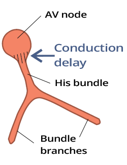
  3. Delayed (decremental) conduction in the AV node
    • The impulse lingers in the AV node for about 0.1s (does not create a curve)
    • Then it moves to His bundle (does not create a curve)
  4. Activation of the ventricular septum
    • From the His bundle the impulse through Purkinje fibers
      • Begins to activate the myocardial septum
      • Starts generating the Q wave

First-Degree AV Block
First degree heart block
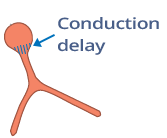
  • The conduction of the impulse through the AV node is prolonged but not blocked
    • It is not a block but a delayed conduction
  • On the ECG, we see a prolonged PQ interval > 0.2s

  • Prolonged impulse conduction can be:
    • Suprahisian (in the AV node - most commonly)
    • Hisian (in the His bundle)
    • Infrahisian (below the His bundle)
    • At the beginning of the bifurcation of the Tawara branches

ECG and First-Degree AV Block


ECG 1st degree AV block, prolonged PR PQ interval

First-Degree AV Block


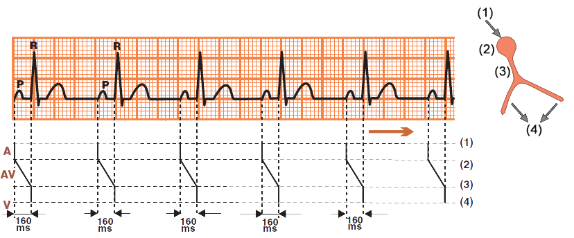
ECG and Laddergram conduction via av node, normal sinus rhythm

Sinus Rhythm

  • Laddergram illustrates the propagation of the impulse through the conduction system
    • A - Atria, AV - AV junction, V - Ventricles
  • PQ interval: 0.16s
    • In an intact AV junction (AV node), the PQ interval is 0.12 - 0.2s


ECG and Laddergram first 1st degree av block

First-Degree AV Block


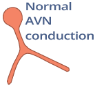
normal av node conduction
ECG normal sinus rhythm, normal conduction via av junction (av node)

Sinus Rhythm


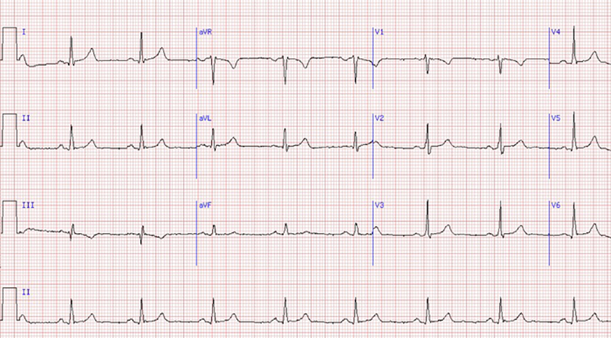
delay av node conduction, first degree av block
ECG first degree av block, prolonged PQ PR interval

First-Degree AV Block


delay av node conduction, first degree av block
ECG first degree heart block, prolonged PQ interval

First-Degree AV Block


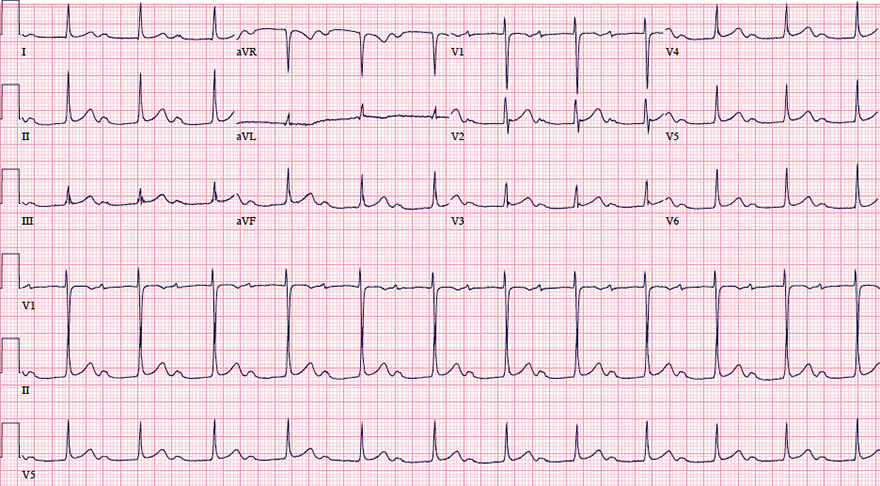
delay av node conduction, first degree av block
ECG first degree av block, sinus bradycardia, prolonged pr interval

First-Degree AV Block and Sinus Bradycardia


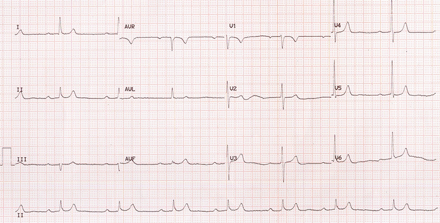
delay av node conduction, first degree av block
ECG sinus bradycardia, av block 1st degree, prolonged PR interval, Beta blocker overdose, toxicity

First-Degree AV Block and Sinus Bradycardia


Sources

  • ECG from Basics to Essentials Step by Step
  • litfl.com
  • ecgwaves.com
  • metealpaslan.com
  • medmastery.com
  • uptodate.com
  • ecgpedia.org
  • wikipedia.org
  • Strong Medicine
  • Understanding Pacemakers


šípka späť

First Degree AV block

First degree AV (heart) block

Atrioventricular (AV) Node

  • In sinus rhythm, impulses are generated regularly (about 60/min.) in the SA node
    • Each impulse spreads through the atria (P wave) to the AV node
  • In the AV node, the impulse slows down by about 0.1s
    • During this time, the atria pump blood into the ventricles
    • Then the impulse continues to the ventricles (QRS complex)

Atrioventricular (AV) node conduction

PQ Interval

  1. Impulse originates in SA node
    • When it moves to the atrial myocardium, it starts generating the P wave
    • Simultaneously, it spreads through the conduction system towards the AV node
      • The impulse in the conduction system does not create a curve
  2. The impulse enters the AV node
    • The impulse spreads from the SA node
    • At the time of atrial activation (peak of the P wave)
      • It arrives through the conduction system to the AV node
  3. Delayed (decremental) conduction in the AV node
    • The impulse lingers in the AV node for about 0.1s (does not create a curve)
    • Then it moves to His bundle (does not create a curve)
  4. Activation of the ventricular septum
    • From the His bundle the impulse through Purkinje fibers
      • Begins to activate the myocardial septum
      • Starts generating the Q wave
AV node conduction, PR interval, PR segment

First-Degree AV Block

  • The conduction of the impulse through the AV node is prolonged but not blocked
    • It is not a block but a delayed conduction
  • On the ECG, we see a prolonged PQ interval > 0.2s

  • Prolonged impulse conduction can be:
    • Suprahisian (in the AV node - most commonly)
    • Hisian (in the His bundle)
    • Infrahisian (below the His bundle)
    • At the beginning of the bifurcation of the Tawara branches

First degree heart block

ECG and First-Degree AV Block


ECG 1st degree AV block, prolonged PR PQ interval

First-Degree AV Block


ECG and Laddergram conduction via av node, normal sinus rhythm

Sinus Rhythm

  • Laddergram illustrates the propagation of the impulse through the conduction system
    • A - Atria, AV - AV junction, V - Ventricles
  • PQ interval: 0.16s
    • In an intact AV junction (AV node), the PQ interval is 0.12 - 0.2s


ECG and Laddergram first 1st degree av block

First-Degree AV Block


ECG normal sinus rhythm, normal conduction via av junction (av node)

Sinus Rhythm

normal av node conduction


ECG first degree av block, prolonged PQ PR interval

First-Degree AV Block

  • Prolonged PQ interval 0.28s (> 0.2s)
    • P wave is embedded within the T wave
  • Sinus Rhythm
delay av node conduction, first degree av block


ECG first degree heart block, prolonged PQ interval

First-Degree AV Block

delay av node conduction, first degree av block


ECG first degree av block, sinus bradycardia, prolonged pr interval

First-Degree AV Block and Sinus Bradycardia

delay av node conduction, first degree av block


ECG sinus bradycardia, av block 1st degree, prolonged PR interval, Beta blocker overdose, toxicity

First-Degree AV Block and Sinus Bradycardia

delay av node conduction, first degree av block


Sources

  • ECG from Basics to Essentials Step by Step
  • litfl.com
  • ecgwaves.com
  • metealpaslan.com
  • medmastery.com
  • uptodate.com
  • ecgpedia.org
  • wikipedia.org
  • Strong Medicine
  • Understanding Pacemakers