ECGbook.com

Making Medical Education Free for All

ECGbook.com

Making Medical Education Free for All

Home /

First Degree SA Block

1st degree Sinoatrial (SA) block

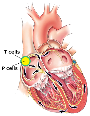
Sinoatrial Node (P and T Cells)

sinus node, P cells, T cells

1st Degree SA Block

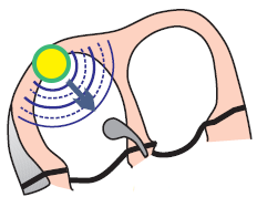
1st degree SA block, Delay between impulse generation and transmission to the atrium

ECG and 1st Degree SA Block

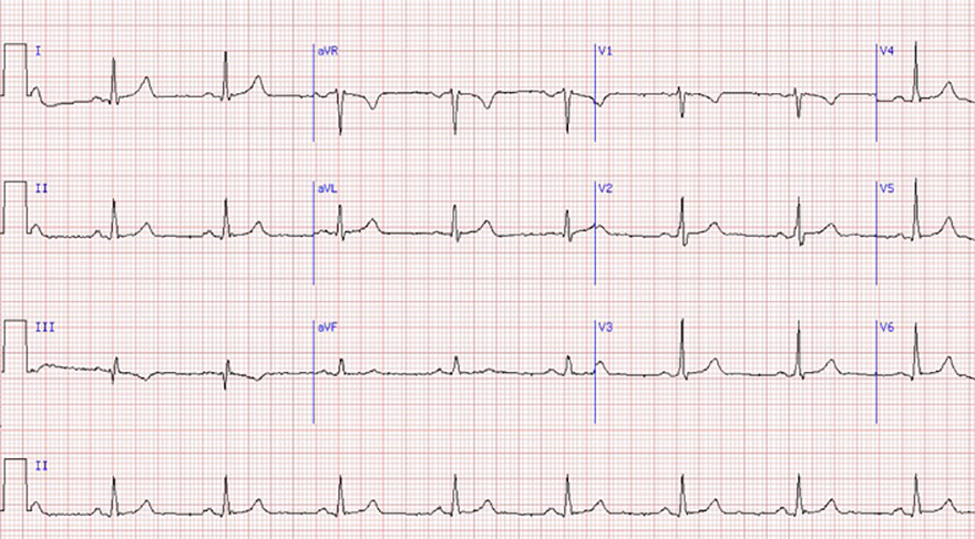
  • The ECG shows a sinus rhythm (physiological P waves are visible)
    • 1st Degree SA Block cannot be distinguished from sinus rhythm

ECG 1st degree SA block, regular P waves

Sinus Rhythm (1st Degree SA Block?)

  • Physiological P waves
  • P waves are a fundamental feature of sinus rhythm
  • The beginning of the P wave occurs when the impulse passes from the SA node (from T cells) to the atria
  • Sinus rhythm and 1st Degree SA Block have the same ECG appearance


Laddergram sinus rhythm

Sinus Rhythm


Laddergram 1st degree SA block

First-Degree SA Block

  • SA node (P cells) generates impulses regularly (green dots)
  • Each impulse quickly reaches the T cells (S-A), where conduction is slowed
    • Conduction in the (S-A) region is prolonged compared to the physiological SA node
    • Then, the impulse from the T cells (S-A) moves to the atrium (A - start of the P wave)
  • PP interval on the ECG is exactly the same as in sinus rhythm
    • It is not possible to distinguish sinus rhythm from first-degree SA block based on the ECG
    • Electrophysiological testing is used for diagnosis


SA node and Sinoatrial first degree block
ECG sinoatrial block, regular sinus P waves

Sinus Rhythm


Sources

  • ECG from Basics to Essentials Step by Step
  • litfl.com
  • ecgwaves.com
  • metealpaslan.com
  • medmastery.com
  • uptodate.com
  • ecgpedia.org
  • wikipedia.org
  • Strong Medicine
  • Understanding Pacemakers


šípka späť

First Degree SA Block

1st degree Sinoatrial (SA) block

Sinoatrial Node (P and T Cells)


sinus node, P cells, T cells

1st Degree SA Block

  • The SA node generates impulses regularly
    • P cells are intact
  • 1st Degree SA Block is prolonged conduction through T cells
    • The impulse passes more slowly (through T cells) from P cells to the atrium
1st degree SA block, Delay between impulse generation and transmission to the atrium

ECG and 1st Degree SA Block

  • The ECG shows a sinus rhythm (physiological P waves are visible)
    • 1st Degree SA Block cannot be distinguished from sinus rhythm

ECG 1st degree SA block, regular P waves

Sinus Rhythm (1st Degree SA Block?)

  • Physiological P waves
  • P waves are a fundamental feature of sinus rhythm
  • The beginning of the P wave occurs when the impulse passes from the SA node (from T cells) to the atria
  • Sinus rhythm and 1st Degree SA Block have the same ECG appearance


Laddergram sinus rhythm

Sinus Rhythm


Laddergram 1st degree SA block

First-Degree SA Block

  • SA node (P cells) generates impulses regularly (green dots)
  • Each impulse quickly reaches the T cells (S-A), where conduction is slowed
    • Conduction in the (S-A) region is prolonged compared to the physiological SA node
    • Then, the impulse from the T cells (S-A) moves to the atrium (A - start of the P wave)
  • PP interval on the ECG is exactly the same as in sinus rhythm
    • It is not possible to distinguish sinus rhythm from first-degree SA block based on the ECG
    • Electrophysiological testing is used for diagnosis


ECG sinoatrial block, regular sinus P waves

Sinus Rhythm


SA node and Sinoatrial first degree block


Sources

  • ECG from Basics to Essentials Step by Step
  • litfl.com
  • ecgwaves.com
  • metealpaslan.com
  • medmastery.com
  • uptodate.com
  • ecgpedia.org
  • wikipedia.org
  • Strong Medicine
  • Understanding Pacemakers