Home /
Second Degree AV Block - High grade
High grade 2nd degree AV block
Atrioventricular (AV) Node

- During sinus rhythm, impulses are generated regularly (about 60/min) in the SA node
- Each impulse spreads through the atria (P wave) to the AV node
- In the AV node, the impulse slows down by about 0.1s
- During this time, the atria pump blood into the ventricles
- Then the impulse continues to the ventricles (QRS complex)
PQ Interval
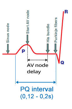
- Impulse originates in the SA node
- When it passes to the atrial myocardium, it begins to generate the P wave
- Simultaneously, it spreads through the conduction system to the AV node
- The impulse in the conduction system does not create a curve
- The impulse enters the AV node
- The impulse spreads from the SA node
- At the time of atrial activation (peak of the P wave)
- It arrives through the conduction system to the AV node
- Slowed (decremental) conduction in the AV node
- The impulse is delayed in the AV node for approx. 0.1s (no curve is created)
- Then it passes into the His bundle (no curve is created)
- Activation of the ventricular septum
- From the His bundle, the impulse travels through the Purkinje fibers
- Begins to activate the myocardium of the ventricular septum
- Begins to generate the Q wave
AV Block II Degree (Mobitz I, Mobitz II)
- Woldemar Mobitz
- He was a Russian physician who worked as a cardiologist in Germany
- In 1924, he described AV block II degree on the ECG and divided it into 2 types (Mobitz I, II)
- Mobitz I (Wenckebach)
- Often referred to as Wenckebach
- Because in the AV node there is the Wenckebach phenomenon
- Mobitz II (Hay)
- John Hay was an English physician who described this AV block II degree based on pulses (without ECG) in 1906
- Later, it was described in more detail by Mobitz and is more commonly referred to as Mobitz II, rarely as Hay

AV Block II Degree - Mobitz I (Wenckebach)
- The defect is in the AV node
- Conduction in the AV node through the AV node gradually lengthens (Wenckebach phenomenon)
- Mobitz I has the Wenckebach phenomenon:
- The PQ interval gradually lengthens
- until the 5th P wave is blocked, then the cycle repeats
- QRS complexes are narrow (<0.12s)
- Conduction to the ventricles is (5:4), from 5 P waves, 4 QRS complexes are produced
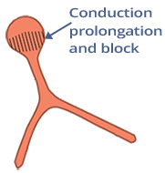
AV Block II Degree, Mobitz II (QRS<0.12s)
- The defect is infranodal (below the AV node)
- The His bundle intermittently blocks impulses (P waves)
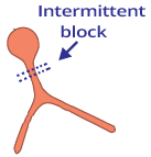
- The PQ interval is constant (Wenckebach phenomenon is not present)
- Mobitz II is an intermittent block in the AV junction (without lengthening of the PQ interval)
- QRS complexes are narrow (<0.12s)
- 25% of Mobitz II AV blocks have narrow QRS complexes
- Because the intermittent block is in the His bundle
- Conduction to the ventricles is (4:3)
AV Block II Degree, Mobitz II (QRS>0.12s)
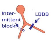
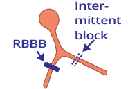
- The defect is infrahisian (below the His bundle)
- The bundle branch or fascicle intermittently blocks impulses (P waves)
- There is a pre-existing bundle branch block (LBBB, RBBB)
- QRS complexes are wide (>0.12s)
- 75% of Mobitz II AV blocks have wide QRS complexes
- because of a pre-existing bundle branch (fascicular) block
- The PQ interval is constant (Wenckebach phenomenon is not present)
- Mobitz II is an intermittent block in the AV junction (without lengthening of the PQ interval)
- Conduction to the ventricles is (3:2)
High-Degree AV Block
- Sinus rhythm has a conduction to the ventricles of 1:1 (P:QRS)
- In second-degree AV block, some P waves are blocked (in the AV junction), so conduction to the ventricles is not 1:1
- Second-degree AV block most commonly has conduction to the ventricles: 3:2, 4:3, 5:4
- Second-degree AV block with conduction to the ventricles (2:1) - can be either Mobitz I or Mobitz II
- If conduction to the ventricles is 3:1 or higher
- it is referred to as a high-degree AV block
- High-degree AV block is almost always of the Mobitz II type
- Hisian has narrow QRS complexes
- Infrahisian has wide QRS complexes
ECG and High-Degree AV Block
- PP interval is regular (SA node generates impulses regularly)
- Conduction to the ventricles is 3:1 or higher (3:1, 4:1, 5:1...)
- PQ interval is constant
- This indicates that the AV node intermittently conducts the P wave to the ventricles (resulting in a QRS)
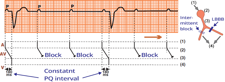
High-Degree AV Block
- Laddergram illustrates the propagation of the impulse through the conduction system
- A - Atria, AV - AV junction, V - Ventricles
- PP interval is regular
- PQ interval is constant (180ms)
- Conduction to the ventricles is 3:1
- This is a second-degree AV block, Mobitz II
- Wide QRS complexes (> 0.12s)
- There is intermittent block of the right bundle branch
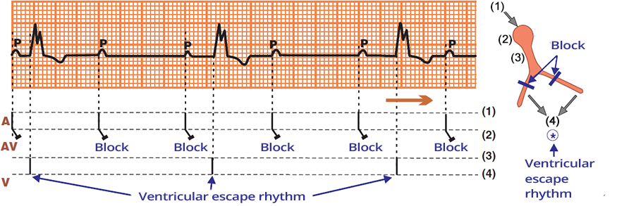
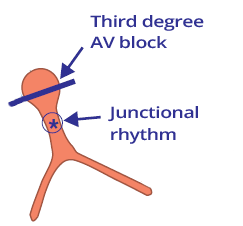
Third-Degree AV Block (Complete AV Block)
- In a third-degree AV block, the AV junction is disrupted
- The atria and ventricles are electrically isolated from each other
- PP interval is regular (the SA node generates impulses regularly - P waves)
- PQ interval changes
- The SA node generates impulses regularly (P waves)
- An activated ventricular focus generates impulses independently of the SA node (wide QRS complexes)
- Independence of the atria (P waves) and ventricles (QRS) is AV dissociation
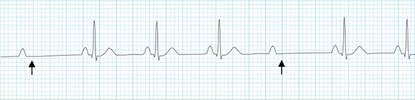
Second-Degree AV Block with 2:1 Conduction
- Conduction to the ventricles is 2:1
- Every second P wave is blocked (blue arrows)
- With 2:1 conduction, it is uncertain whether it is Mobitz I or Mobitz II?
- It is unclear whether the PQ interval is prolonged (Mobitz I) or if every 2nd P wave is blocked (Mobitz II)
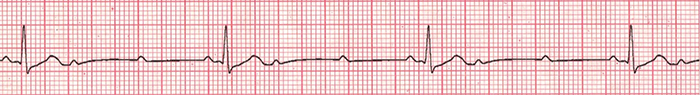
High-Grade AV Block (3:1)
High-Grade AV Block
- PP interval is constant
- PQ interval is constant at 0.2s
- Conduction to the ventricles is 4:1
- This indicates a High-Grade AV Block
- This is a Hisian second-degree AV block - Mobitz II

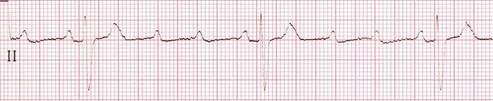
Third-Degree AV Block (Complete AV Block)
- PP interval is constant
- PQ interval varies
- The SA node generates impulses regularly (P waves)
- A secondary pacemaker has activated in the AV junction
- Third-Degree AV Block
- Conduction through the AV junction is interrupted
- The atria (P waves) and ventricles (QRS) are independent of each other (AV dissociation)
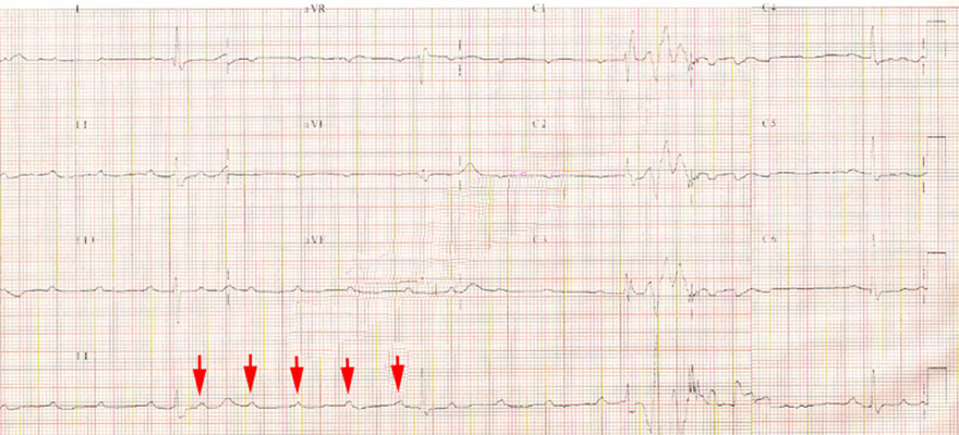
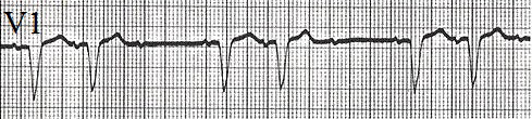
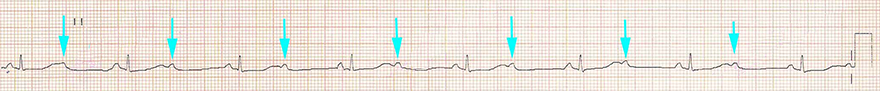
High-Grade AV Block (5:1)
- PP interval is constant
- PQ interval is constant 0.2s
- Conduction to the ventricles is 5:1
- Out of five P waves, one is conducted to the ventricles (blue arrow)
- This is a High-Grade AV Block
- This is a Infra-Hisian AV Block II Degree - Mobitz II
- QRS complexes are wide (>0.12s)
- And have the shape of RBBB — note the V1 lead
Sources
- ECG from Basics to Essentials Step by Step
- litfl.com
- ecgwaves.com
- metealpaslan.com
- medmastery.com
- uptodate.com
- ecgpedia.org
- wikipedia.org
- Strong Medicine
- Understanding Pacemakers