Home /
Second Degree AV Block - Mobitz I (Wenckebach)
2nd degree AV Block, Mobitz I (Wenckebach)
Atrioventricular (AV) Node

- In sinus rhythm, impulses are generated regularly (approximately 60/min.) in the SA node
- Each impulse spreads through the atria (P wave) to the AV node
- The impulse slows down in the AV node by approximately 0.1s
- During this time, the atria pump blood into the ventricles
- Then the impulse continues to the ventricles (QRS complex)
PQ Interval
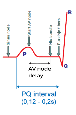
- Impulse originates in the SA node
- When it travels to the atrial myocardium, the P wave begins to form
- Simultaneously, it spreads through the conduction system towards the AV node
- The impulse in the conduction system does not create a curve
- The impulse enters the AV node
- The impulse spreads from the SA node
- At the time of atrial activation (peak of the P wave)
- It arrives through the conduction system to the AV node
- Delayed (decremental) conduction in the AV node
- The impulse lingers in the AV node for about 0.1s (no curve is generated)
- Then it moves into the His bundle (no curve is generated)
- Activation of the ventricular septum
- From the His bundle the impulse through Purkinje fibers
- Begins to activate the myocardial tissue of the ventricular septum
- Begins to form the Q wave
Wenckebach Phenomenon
- Wenckebach phenomenon is a disorder of the conduction system
- in which the conduction of impulses progressively lengthens through a certain part of the conduction system
- until the impulse is blocked, and the cycle then repeats
- It is a cyclic pathological progression of decremental conduction
- The phenomenon was described by the Dutch internist Karel Frederik Wenckebach without an ECG
- He examined the pulse on the radial artery of a woman who complained of irregular pulse
- SA Block II Degree, Type I (Wenckebach)
- It is the progressive lengthening of conduction through the SA node (T cells)
- AV Block II Degree, Mobitz I (Wenckebach)
- It is the progressive lengthening of conduction through the AV node
AV Block II Degree - Mobitz I (Wenckebach)
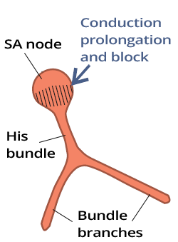
- Conduction of each subsequent impulse
- gradually lengthens through the AV node
- until the impulse is blocked in the AV node
- The cycle then repeats
- On the ECG, the PQ interval progressively lengthens
- Until after the P wave, no QRS complex follows
- The cycle then repeats
- In AV node dysfunction
- The Wenckebach phenomenon occurs in the AV node
- Conduction gradually lengthens until it is blocked - Mobitz I
- In infra-nodal dysfunction (His bundle)
- The Wenckebach phenomenon does not occur
- The "all or nothing" law applies
- The impulse is either conducted or blocked
- Blockage of all impulses is AV Block III Degree
ECG and AV Block II Degree - Mobitz I (Wenckebach)
- PP interval is always the same
- Because the SA node generates impulses regularly (sinus rhythm)
- PQ interval progressively lengthens
- Gradually, the AV node "fatigues" until the P wave is not conducted (after P wave, no QRS complex follows)
- PQ interval after RR pause is shorter than PQ interval before the pause

AV Block II Degree, Mobitz I (Wenckebach)
- PP interval is always the same (sinus rhythm)
- PQ interval progressively lengthens (Wenckebach phenomenon)
- After the 4th P wave, no QRS complex follows
- PQ interval after the pause is shorter than PQ interval before the pause
- Conduction to the ventricles is (4:3)
- Out of 4 P waves, 3 P waves are conducted to the ventricles
- Since there are 3 QRS complexes, the cycle then repeats

Sinus Rhythm
- Laddergram illustrates the spread of the impulse through the conduction system
- A - Atria, AV - AV junction, V - Ventricles
- PQ interval: 0.16s
- With a normal AV junction (AV node), the PQ interval is 0.12 - 0.2s

Second-Degree AV Block - Mobitz I (Wenckebach)
- PP interval is constant: 810ms (sinus rhythm)
- PQ interval progressively lengthens until the P wave is not conducted
- 1st PQ (190ms)
- 2nd PQ (290ms)
- 3rd P wave is blocked
- The cycle then repeats
- Conduction to the ventricles is (3:2)
- Out of 3 P waves, 2 P waves are conducted to the ventricles (resulting in 2 QRS complexes), and the cycle then repeats
Second-Degree AV Block (Mobitz I, Mobitz II)
- Woldemar Mobitz
- He was a Russian physician who worked as a cardiologist in Germany
- In 1924, he described second-degree AV block on an ECG and divided it into 2 types (Mobitz I, II)
- Mobitz I (Wenckebach)
- It is often referred to as Wenckebach
- Because the Wenckebach phenomenon occurs in the AV node
- Mobitz II (Hay)
- John Hay was an English physician who, in 1906, described this second-degree AV block based on pulse (without ECG)
- Later, Mobitz described it in more detail and it is more commonly referred to as Mobitz II, rarely as Hay
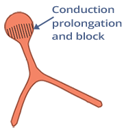

Second-Degree AV Block (Mobitz I)
- Disorder is in the AV node
- Mobitz I features the Wenckebach phenomenon:
- PQ interval progressively prolongs
- Until the 5th P wave is blocked, then the cycle repeats
- Conduction to the ventricles is (5:4)
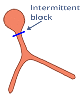

Second-Degree AV Block (Mobitz II)
- Disorder is infranodal (below the AV node)
- PQ interval remains constant (Wenckebach phenomenon is not present)
- AV Block II Degree, Mobitz II
- Any P waves are intermittently blocked (without prolongation of the PQ interval)
- The infranodal area of the AV junction intermittently blocks impulses
- Conduction to the ventricles is (4:3)
Conduction to the Ventricles
- Sinus Rhythm has conduction to the ventricles 1:1 (P:QRS)
- Each P wave is followed by a QRS complex
- In second-degree AV block, some P waves are blocked (in the AV node)
- Therefore, conduction to the ventricles is not 1:1
- Second-Degree AV Block - Mobitz I commonly has conduction to the ventricles as: 3:2, 4:3, 5:4
- Variable conduction to the ventricles means that the conduction to the ventricles changes on the ECG, for example:
- Second-Degree AV Block - Mobitz I with variable conduction to the ventricles (3:2) and (4:3)
- Second-Degree AV Block with Conduction to the Ventricles (2:1)
- Could it be Mobitz I or Mobitz II?


Second-Degree AV Block - Mobitz I (Wenckebach)
- PP interval is constant
- PQ interval gradually lengthens
- Until the P wave is blocked
- The cycle then repeats
- Conduction to the ventricles is 5:4


Second-Degree AV Block - Mobitz I (Wenckebach)
- PP interval is constant
- PQ interval gradually lengthens
- Until the P wave is blocked
- The cycle then repeats
- Variable conduction to the ventricles (5:4) and (6:5)

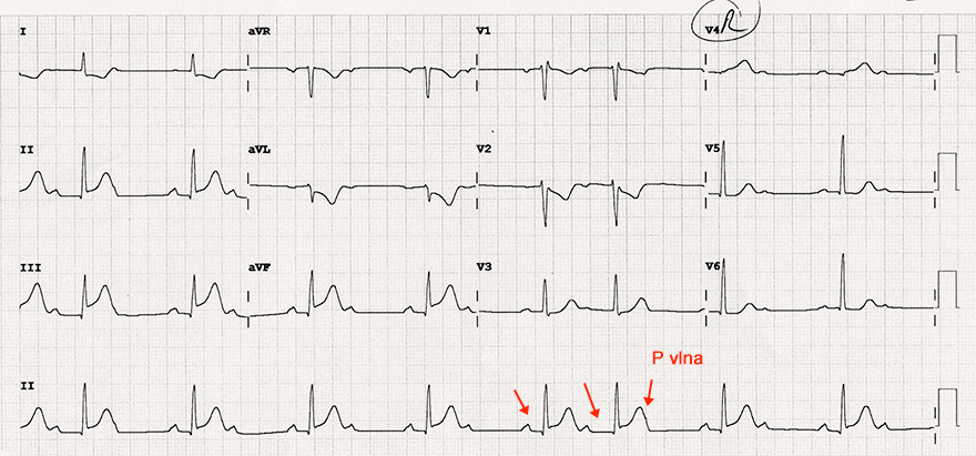
Second-Degree AV Block - Mobitz I (Wenckebach) and Inferior Wall STEMI


Second-Degree AV Block - Mobitz I (Wenckebach)
- Laddergram illustrates the propagation of impulses through the conduction system
- A - Atrium, AV - AV junction, V - Ventricles
- PP interval is constant
- PQ interval progressively prolongs
- Until the P wave is blocked
- The cycle then repeats
- Conduction to the ventricles is (5:4)


Second-Degree AV Block - Mobitz I (Wenckebach)
- PP interval is constant (800ms)
- PQ interval progressively prolongs (160ms...320ms)
- Until the P wave is blocked
- The cycle then repeats
- Conduction to the ventricles is (6:5)


Atypical AV Block II. Degree - Mobitz I (Wenckebach)
- On the ECG, there is a continuous ECG record (in 2 lines)
- PP interval is constant (800ms)
- PQ interval prolongs irregularly
- Until the P wave is blocked
- The cycle then repeats
- Conduction to the ventricles is irregular
- PQ interval before the blocked P wave is longer
- than PQ interval after the blocked P wave
- It is Atypical AV Block II. Degree - Mobitz I (Wenckebach)
- Because PQ interval prolongs irregularly


AV Block II. Degree - Mobitz I (Wenckebach)
- On the ECG, there is a continuous ECG record (in 2 lines)
- PP interval is constant (800ms)
- PQ interval prolongs irregularly
- Some PQ intervals remain the same and do not prolong
- P wave is blocked
- The cycle then repeats
- Conduction to the ventricles is irregular
- PQ interval before the blocked P wave is longer
- than PQ interval after the blocked P wave
- It is Atypical AV Block II. Degree - Mobitz I (Wenckebach)
- Because PQ interval prolongs irregularly


- PP interval is constant (800ms)
- PQ interval is constant (190ms)
- PQ interval after a blocked P wave IS NOT shorter
- than PQ interval before the blocked P wave
- It is AV Block II. Degree - Mobitz II
- Because PQ interval REMAINS constant, even after a blocked P wave


AV Block II. Degree - Mobitz I (Wenckebach)
- PP interval is constant (800ms)
- PQ interval is constant (190ms)
- Except for the PQ interval after a blocked P wave (160ms)
- PQ interval after a blocked P wave IS shorter
- than PQ interval before the blocked P wave
- PQ interval after a blocked P wave (160ms) is shorter
- by 30ms compared to PQ before the blocked P wave
- It is AV Block II. Degree - Mobitz I (Wenckebach)


AV Block II. Degree - Mobitz I (Wenckebach)
- PP interval is constant (800ms)
- PQ interval is constant (190ms)
- except for the PQ interval after a blocked P wave (175ms)
- PQ interval after a blocked P wave IS shorter
- than PQ interval before the blocked P wave
- PQ interval after a blocked P wave (175ms) is shorter
- by 15ms compared to PQ before the blocked P wave
- Some criteria have a 20ms tolerance
- and might classify this AV block as Mobitz II
- It is AV Block II. Degree - Mobitz I (Wenckebach)


AV Block II. Degree - Mobitz I (Wenckebach)
- PP interval is constant
- PQ interval is constant
- However, PQ interval after a blocked P wave is shorter
- Without a laddergram, it is harder to evaluate
- It is AV Block II. Degree - Mobitz I (Wenckebach)
- If the PQ interval after the blocked P wave were the same
- as before the blocked P wave, it would be classified as Mobitz II


AV Block II. Degree - Non-Classifiable
- PP interval is constant
- PQ interval is constant
- On the ECG, the PQ interval after the blocked P wave is not visible
- If the PQ interval after the blocked P wave were:
- Shortened - it would be Mobitz I
- Constant - it would be Mobitz II
Sources
- ECG from Basics to Essentials Step by Step
- litfl.com
- ecgwaves.com
- metealpaslan.com
- medmastery.com
- uptodate.com
- ecgpedia.org
- wikipedia.org
- Strong Medicine
- Understanding Pacemakers


