Home /
Second Degree SA Block - Type I (Wenckebach)
2nd degree Sinoatrial (SA) block, type I (Wenckebach)
SA Node (P and T Cells)

Wenckebach Phenomenon
- The Wenckebach phenomenon is a disorder of the conduction system
- where there is a gradual prolongation of impulse conduction through a specific part of the conduction system
- until the impulse is blocked, and then the cycle repeats
- It is a cyclic pathological progression of decremental conduction
- The phenomenon was described by the Dutch internist Karel Frederik Wenckebach without ECG
- He examined the pulse on the radial artery of a woman who complained of irregular pulse
- SA Block II Degree, Type I (Wenckebach)
- There is a gradual prolongation of conduction through the SA node (T cells)
- AV Block II Degree, Mobitz I (Wenckebach)
- There is a gradual prolongation of conduction through the AV node
SA Block II Degree - Type I (Wenckebach)
- The SA node generates impulses regularly
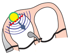
- Upon impulse exit from the SA node (in T cells), the Wenckebach phenomenon occurs
- Impulse conduction at the exit from the SA node progressively lengthens
- until the impulse is blocked, and the cycle repeats
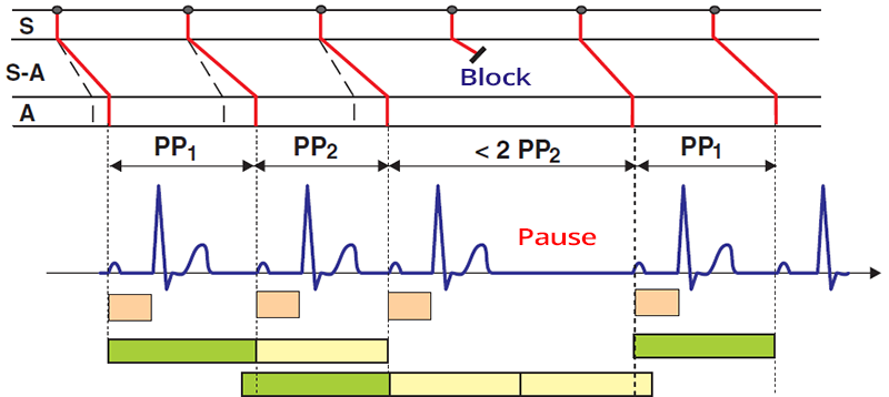
ECG and SA Block II Degree - Type I (Wenckebach)
- The ECG shows a sinus rhythm (we see physiological P waves)
- PP interval progressively shortens until a pause follows
- The pause is shorter than twice the shortest PP (2PP) interval
- PQ interval remains unchanged (AV node is intact)

SA Block II Degree - Type I (Wenckebach)
- PP interval progressively shortens from 740ms to 700ms
- Then the impulse from the SA node is blocked and is followed by a pause of 1300ms
- PP interval with the pause (1300) is shorter than twice the shortest PP interval (2 x 700 = 1400)
- PQ interval is constant (because the AV node is intact)

Sinus Rhythm
- Laddergram illustrates the propagation of impulses through the conduction system
- S - P cells (SA node), S-A - T cells (Sinoatrial junction), A - atrium
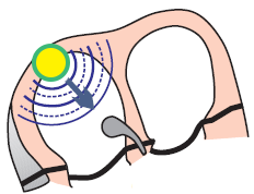
- In sinus rhythm, the SA node (P cells) generates impulses regularly (green dots)
- Each impulse quickly reaches the T cells (S-A), where conduction slows down
- Then, the impulse from the T cells (S-A) moves to the atrium (A - start of the P wave)
- PP interval is consistent on the ECG

SA Block II. Degree - Type I (Wenckebach)
- Laddergram: S - P cells (SA node), S-A - T cells (Sinoatrial junction), A - atrium
- SA node (P cells) generates impulses regularly (green dots)
- In T cells - SA junction (S-A), the conduction of impulses from the SA node to the atrium gradually lengthens
- Therefore, the PP interval shortens (PP1 > PP2)
- Then a pause follows, which is shorter than twice the shortest PP interval
- PP2 pause < 2PP2
- During the pause, the SA node (T cells) "rests," so the subsequent impulse is conducted faster


Sinus Rhythm


SA Block II. Degree, Type I (Wenckebach)
- PP interval shortens (1000ms...820ms)
- PP interval with a dropped P wave (1580ms) is shorter
- than twice the shortest PP interval (2 x 820 = 1640ms)
- PQ interval is constant


SA Block II. Degree - Type II
- PP interval is constant (970ms)
- PP interval with a dropped P wave (1940ms) is equal
- to twice the PP interval without a pause (2 x 970 = 1940)
- PQ interval is constant
- SA Block II. Degree - Type II
- SA node generates impulses regularly
- Impulse conduction from the SA node does not prolong
- Impulses from the SA node intermittently block (PP pause)


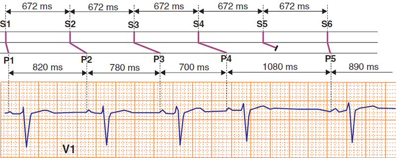
SA Block II. Degree, Type I (Wenckebach)
- Laddergram
- SA node generates impulses regularly S1-S6 (672ms)
- Impulse conduction to the atrium through the SA junction gradually prolongs
- PP intervals shorten P1-P4 (820...700ms)
- A PP pause (1080ms) occurs, which is shorter
- than twice the shortest PP interval
(2 x 700 = 1400ms)
Sources
- ECG from Basics to Essentials Step by Step
- litfl.com
- ecgwaves.com
- metealpaslan.com
- medmastery.com
- uptodate.com
- ecgpedia.org
- wikipedia.org
- Strong Medicine
- Understanding Pacemakers


