|
|
ECGbook.com Making Medical Education Free for All |
|
|
ECGbook.com Making Medical Education Free for All |
|
|
ECGbook.com Making Medical Education Free for All |
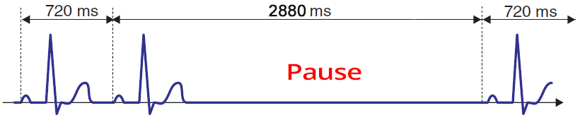
Third-Degree SA Block
Third-Degree SA Block and Sinus Rhythm
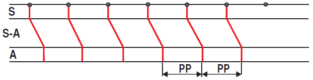
Sinus Rhythm
Third-Degree SA Block
Sinoatrial Rhythm
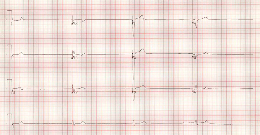
Third-Degree SA Block
Second-Degree SA Block - Type II
Third-Degree SA Block
Sources
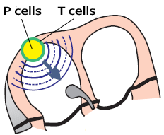
SA Node (P and T Cells)
|
|
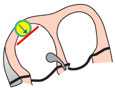
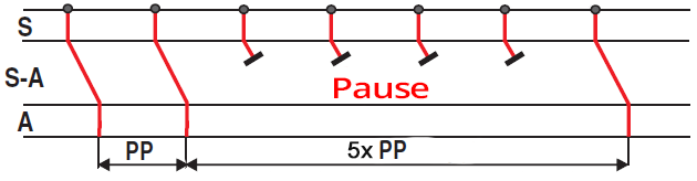
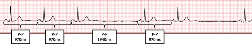
SA Block III Degree
|
|
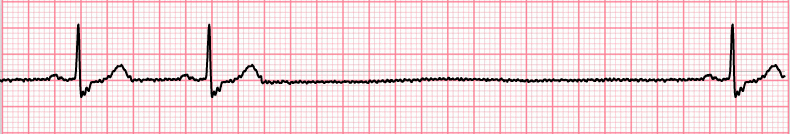
Third-Degree SA Block
Third-Degree SA Block and Sinus Rhythm
Sinus Rhythm
Third-Degree SA Block
|
Sinoatrial Rhythm
|
|
|
Third-Degree SA Block
|
|
|
Second-Degree SA Block - Type II
|
|
|
Third-Degree SA Block
|
|
Sources