|
|
ECGbook.com Making Medical Education Free for All |
|
|
ECGbook.com Making Medical Education Free for All |
|
|
ECGbook.com Making Medical Education Free for All |
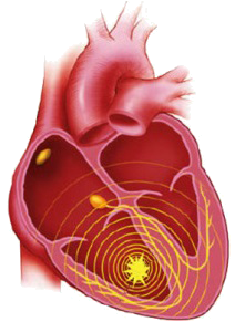
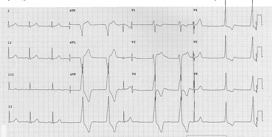
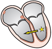
Accelerated Ventricular Rhythm
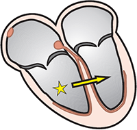
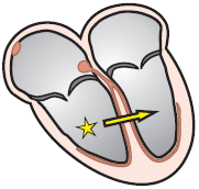
Ventricular Rhythm

Accelerated Ventricular Rhythm
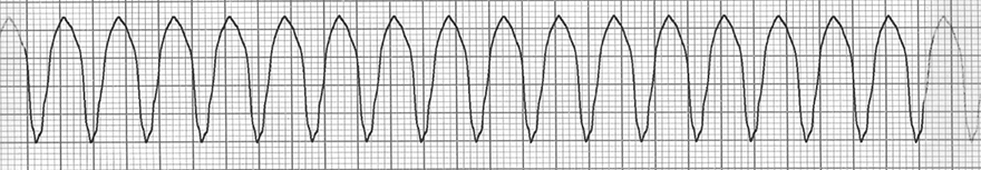
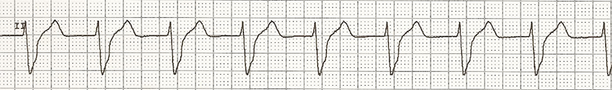
Ventricular Tachycardia
Accelerated Ventricular Rhythm
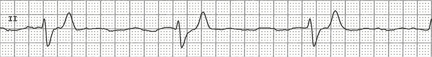
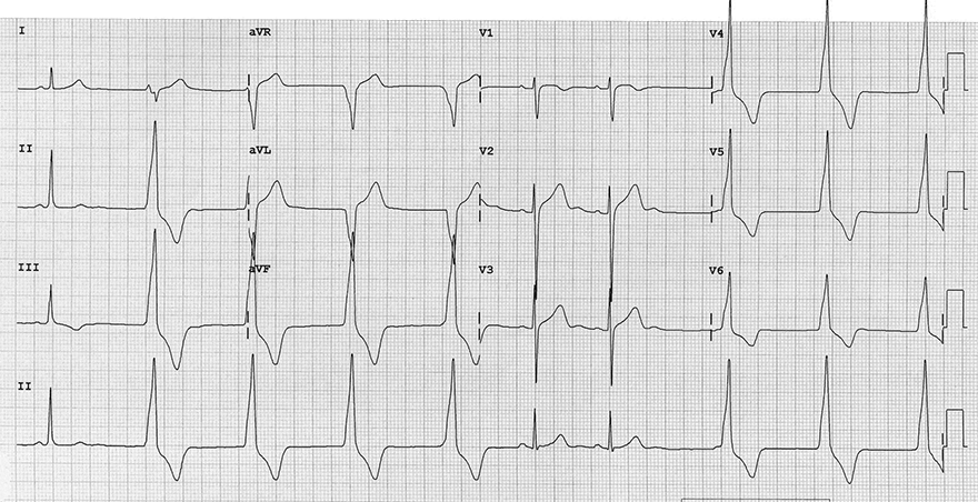
Sinus Rhythm and Ventricular Extrasystoles
Accelerated Ventricular Rhythm
Accelerated Ventricular Rhythm
Sources
Ventricular Rhythm
|
|
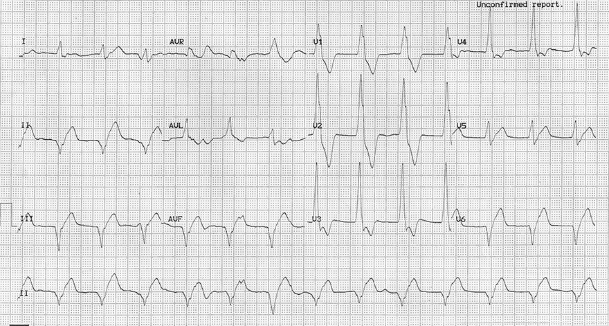
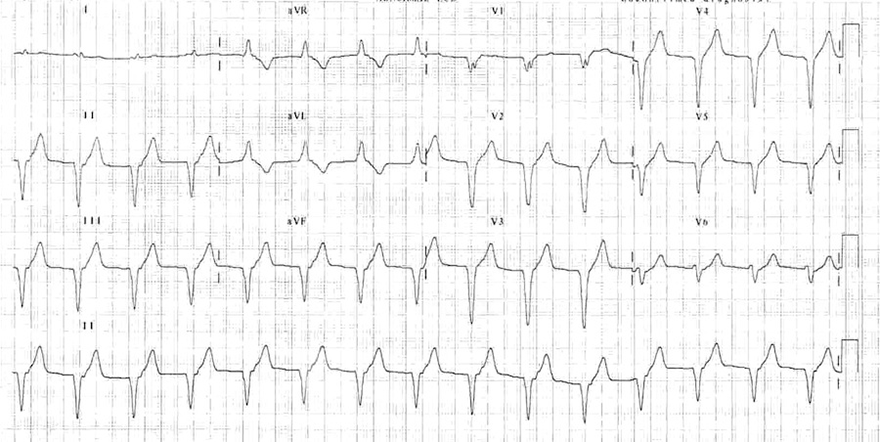
ECG and Accelerated Ventricular Rhythm
|
|
Accelerated Ventricular Rhythm
Ventricular Rhythm
Accelerated Ventricular Rhythm
Ventricular Tachycardia
|
Accelerated Ventricular Rhythm
|
|
|
Sinus Rhythm and Ventricular Extrasystoles
|
|
|
Accelerated Ventricular Rhythm
|
|
|
Accelerated Ventricular Rhythm
|
|
Sources