|
|
ECGbook.com Making Medical Education Free for All |
Upload ECG for Interpretation |
|
|
ECGbook.com Making Medical Education Free for All |
Upload ECG for Interpretation |
|
|
ECGbook.com Making Medical Education Free for All |
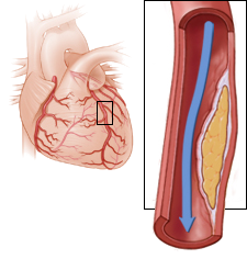
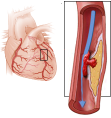
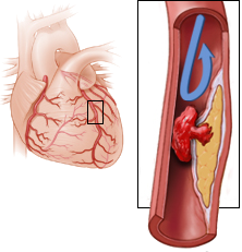
Atherosclerosis and Acute Coronary Syndrome
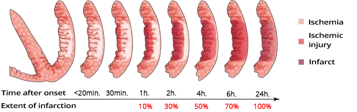
Dynamics of Ischemia after Occlusion
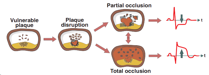
ST Segment and Acute Coronary Syndrome
Subendocardial Ischemia

Subepicardial Ischemia

Acute Coronary Syndrome
STEMI Infarction

NSTEMI Infarction
Ischemia Post-Ergometry

Unstable Angina Pectoris
Acute STEMI of the Anterior Wall
Sources
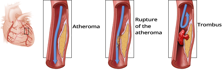
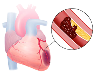
Atherosclerosis and Acute Coronary Syndrome
Dynamics of Ischemia in Occlusion
|
|
Dynamics of Ischemia after Occlusion
ST Segment and Acute Coronary Syndrome
|
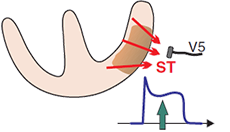
Subendocardial Ischemia
|
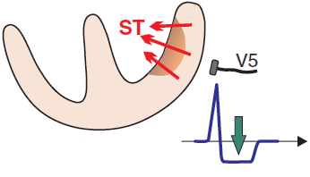
Subepicardial Ischemia
|
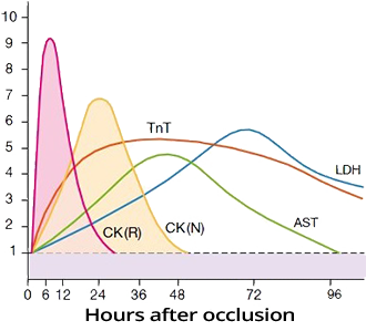
Troponin and Infarction
|
|
Acute Coronary Syndrome (ACS)
|
|
Acute Coronary Syndrome
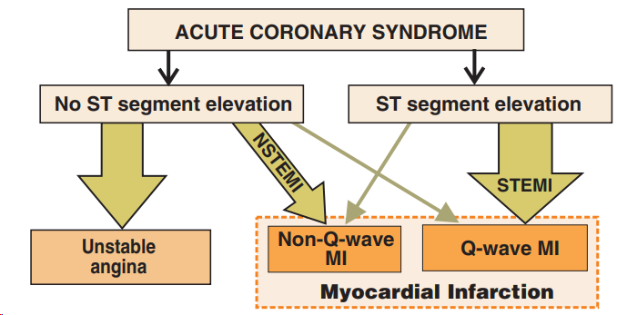
Nomenclature of Infarction
|
STEMI Infarction
|
NSTEMI Infarction
|
|
Ischemia Post-Ergometry
|
|
|
Unstable Angina Pectoris
|
|
|
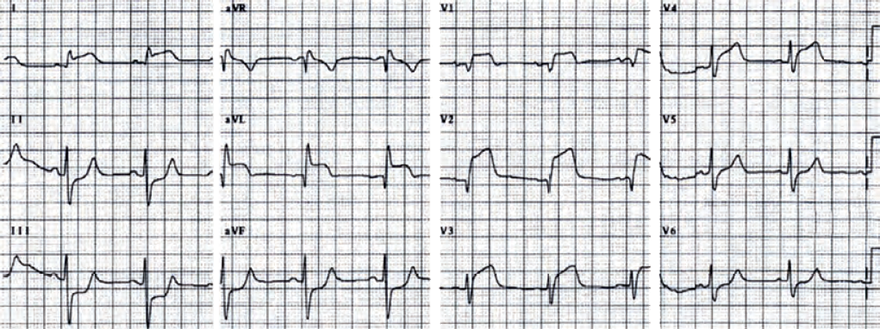
Acute STEMI of the Anterior Wall
|
|
Sources