ECGbook.com

Making Medical Education Free for All

ECGbook.com

Making Medical Education Free for All

Home /

Action Potential

Action potential

Working Myocardium

Heart muscle and syncytium, intercalated discs, gap junctions

Cardiomyocytes of the working myocardium
  • Cardiomyocytes in working myocardium have cylindrical (elongated) shape
  • They are branched, with 2 or more branches at the ends, which connect to other cardiomyocytes
  • They are interconnected by intercalated discs
    • The intercalated discs contain gap junctions (nexus) through which ions can freely pass
    • Thus, action potential spreads very quickly through cardiomyocytes
    • Cardiomyocytes form a syncytium
      • It is a multi-cellular structure formed by fusion of many cells
      • In principle, it is a multi-cellular structure with shared cytoplasm
  • They contain troponin, which is released upon their damage - necrosis (in case of myocardial infarction)


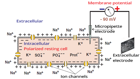
Polarized resting cell with intracellular and extracellular electrode. Sodium potassium pump with ions channels

Cardiomyocyte and Resting Membrane Potential


Action Potential

  • It is a rapid change in electrical voltage of the cardiomyocyte
  • It arises due to changes in concentration of intracellular and extracellular ions
    • Action potential propagates more rapidly through conduction system than through working myocardium
  • It involves depolarization - reversal of membrane polarity
  • Subsequently, repolarization occurs - returning to the original state


Resting membrane potential with -90mV

Resting Membrane Potential


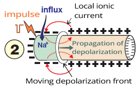
Propagation of depolarization, electrical impulse, sodium influx, and local ionic current

Depolarization

  • When the cardiomyocyte is stimulated by a negative electrical impulse
  • This opens Na+ ion channels
    • Followed by influx of Na+ into the cell due to the electrical gradient
    • Causing a change in electrical voltage
      • because intracellular Na+ concentration increases
  • When the membrane potential reaches the threshold potential (-50mV)
    • additional rapid sodium channels open
  • The result is depolarization
    • It involves a reversal of membrane polarity
    • The extracellular space becomes negative
      • The intracellular space becomes positive due to Na+ influx
    • Depolarization spreads from the activation site as a depolarization wave


Depolarized cell with negative extracellular space

Depolarized Cardiomyocyte


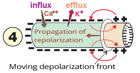
Propagation of repolarization, influx calcium, efflux potassium

Repolarization

  • At the end of depolarization, there is a movement of ions through channels again
    • Mainly involving Ca++ and K+ ions
  • This results in a return to the original polarized state
  • The repolarization wave spreads slowly and oppositely to depolarization
    • In the myocardium, epicardium begins repolarizing earlier than endocardium
  • The extracellular environment again changes to positive


Resting cell after repolarization

Resting Membrane Potential

Propagation of Action Potential

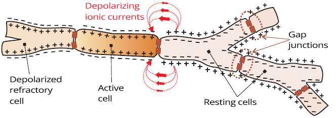
Longitudinal propagation action potential with depolarization wave, syncytium, intercalated discs and gap junctions

Cardiomyocytes of the Working Myocardium

  • They are branched and longitudinally connected by intercalated discs
  • The cytoplasm of cardiomyocytes is connected through intercalated discs
    • The entire muscle fiber behaves like a single long cell (syncytium)
  • The depolarization wave propagates throughout the myocardium as if through a single cell


Longitudinal and transversal propagation wave of action potential

Working Myocardium


Action Potential and Electrical Voltage

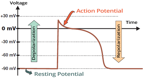
Action potential with depolarization and repolarization voltage
  • Volt is the difference in electrical potential between 2 points
    • The 1st point is the intracellular environment
    • The 2nd point is the extracellular environment
  • Resting Membrane Potential
    • Has a value of -90mV
    • The cell is negative intracellularly
  • Depolarized Cardiomyocyte
    • Has a value of 15mV
    • The cell is positive intracellularly
  • During the action potential
    • There is a change in ion distribution
    • And consequently, in electrical voltage

Action Potential and Contraction of Cardiomyocyte


Action Potential in the Conduction System

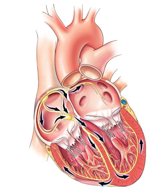
Heart conduction system and action potential propagation
  • Action potential (impulse) originates spontaneously in the SA node
  • From the SA node, it begins to activate the atrial myocardium
    • Spreads like throwing a stone into water
      • From the SA node diffuses into the atria (P wave)
    • Resulting in atrial systole
  • The impulse simultaneously rapidly spreads through the conduction system to the AV node
    • From the AV node to the Bundle of His and to the ventricles
    • First activates the septum (Q wave), and then the ventricles (R wave)

  • Conduction system
    • Spontaneously generates impulses in the SA node
    • Does not contract
    • Conducts impulse rapidly
    • The impulse spreads from the conduction system to the working myocardium
  • Working myocardium
    • Does not spontaneously generate impulses
    • Contracts
      • Each impulse triggers systole
    • Conducts impulse more slowly than the conduction system

Action Potential in the SA Node

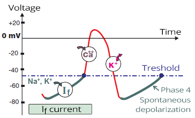
SA node action potential and If (funny current)

If Channels and Heart Rate

  • If channels in the SA node spontaneously depolarize and determine the heart rate
  • Their depolarization rate is influenced by the autonomic nervous system
    • Activation of the sympathetic system releases catecholamines (adrenaline, noradrenaline)
    • The depolarization rate increases if the slope during the If current is steeper
      • This allows the curve to reach the threshold value (-50mV) sooner


Sa node cycle shortening via funny current

Stimulation of the SA Node


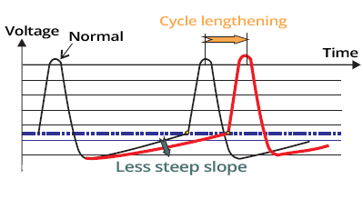
SA node depolarization cycle lengthening, if funny current

Inhibition of the SA Node

  • For example, certain medications
  • The If current curve of the action potential becomes flatter
  • The 4th phase of the action potential lengthens
  • The heart rate decreases

Working Myocardium vs. SA Node


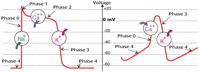
Phases of action potential in conduction system (SA node) and ventricular myocyte

Action potential in
working myocardium

Action potential in
SA node


Heart areas with action potentials in conduction system and myocardial muscle

Action Potential in Various Parts of the Heart
  • Action potential in different parts of the heart has a different shape
    • It varies especially in the conduction system and working myocardium
    • Due to distinct properties of cardiomyocytes
  • Action potential (impulse) of the working myocardium is captured by an ECG device and results in an ECG waveform

Suppression of the Conduction System

Dominant pacemaker - SA node and overdrive suppression

ECG and Action Potential

Action potential duration and one electrical cardiac cycle

ECG (lead V6) and Action Potential

  • QRS complex represents ventricular depolarization
    • During the QRS complex, a depolarization wave spreads through the ventricles
  • ST segment is an isoelectric line
  • T wave represents ventricular repolarization
    • The electrical vector during repolarization has the same direction as during depolarization
    • However, repolarization takes longer, hence the T wave is wider than the QRS complex
  • QT interval corresponds to 1 electrical cardiac cycle:
    • Ventricular depolarization + repolarization
  • P wave represents atrial depolarization
    • The action potential of the P wave is not shown in the image
    • Atrial repolarization is hidden within the QRS complex

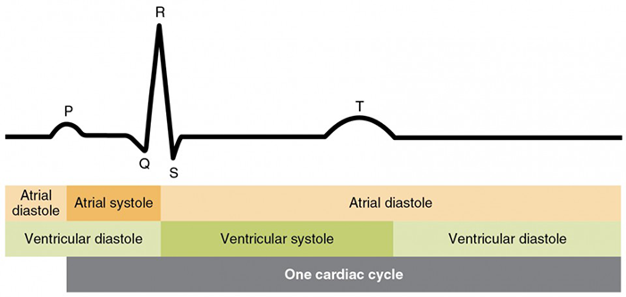
ECG and Cardiac Cycle

action potential, atrial diastole, atrial systole, ventricular diastole, ventricular systole, one cardiac cycle

ECG and Cardiac Cycle


Sources

  • ECG from Basics to Essentials Step by Step
  • litfl.com
  • ecgwaves.com
  • metealpaslan.com
  • medmastery.com
  • uptodate.com
  • ecgpedia.org
  • wikipedia.org
  • Strong Medicine
  • Understanding Pacemakers


šípka späť

Action Potential

Action potential

Working Myocardium

Heart muscle and syncytium, intercalated discs, gap junctions

Cardiomyocytes of the working myocardium

  • Cardiomyocytes in working myocardium have cylindrical (elongated) shape
  • They are branched, with 2 or more branches at the ends, which connect to other cardiomyocytes
  • They are interconnected by intercalated discs
    • The intercalated discs contain gap junctions (nexus) through which ions can freely pass
    • Thus, action potential spreads very quickly through cardiomyocytes
    • Cardiomyocytes form a syncytium
      • It is a multi-cellular structure formed by fusion of many cells
      • In principle, it is a multi-cellular structure with shared cytoplasm
  • They contain troponin, which is released upon their damage - necrosis (in case of myocardial infarction)


Polarized resting cell with intracellular and extracellular electrode. Sodium potassium pump with ions channels

Cardiomyocyte and Resting Membrane Potential


Action Potential

  • It is a rapid change in electrical voltage of the cardiomyocyte
  • It arises due to changes in concentration of intracellular and extracellular ions
    • Action potential propagates more rapidly through conduction system than through working myocardium
  • It involves depolarization - reversal of membrane polarity
  • Subsequently, repolarization occurs - returning to the original state


Resting membrane potential with -90mV

Resting Membrane Potential

  • During rest, the cardiomyocyte is in a polarized state
  • The extracellular space is electrically positive
    • Due to the high extracellular concentration of Na+
  • The intracellular space is negative
  • The voltage difference (intra vs extracellular) is -90mV


Propagation of depolarization, electrical impulse, sodium influx, and local ionic current

Depolarization

  • When the cardiomyocyte is stimulated by a negative electrical impulse
  • This opens Na+ ion channels
    • Followed by influx of Na+ into the cell due to the electrical gradient
    • Causing a change in electrical voltage
      • because intracellular Na+ concentration increases
  • When the membrane potential reaches the threshold potential (-50mV)
    • additional rapid sodium channels open
  • The result is depolarization
    • It involves a reversal of membrane polarity
    • The extracellular space becomes negative
      • The intracellular space becomes positive due to Na+ influx
    • Depolarization spreads from the activation site as a depolarization wave


Depolarized cell with negative extracellular space

Depolarized Cardiomyocyte

  • The extracellular environment is negative
  • The depolarization wave gradually depolarizes the entire cardiomyocyte
    • Then it continues to adjacent cardiomyocytes


Propagation of repolarization, influx calcium, efflux potassium

Repolarization

  • At the end of depolarization, there is a movement of ions through channels again
    • Mainly involving Ca++ and K+ ions
  • This results in a return to the original polarized state
  • The repolarization wave spreads slowly and oppositely to depolarization
    • In the myocardium, epicardium begins repolarizing earlier than endocardium
  • The extracellular environment again changes to positive


Resting cell after repolarization

Resting Membrane Potential

  • During the action potential, specific channels open at a specific time
    • Ion transfer is passive following the electrical gradient
  • At the end of the cycle, mainly the Na+/K+ pump redistributes ions to their original state

Propagation of Action Potential

Longitudinal propagation action potential with depolarization wave, syncytium, intercalated discs and gap junctions

Cardiomyocytes of the Working Myocardium

  • They are branched and longitudinally connected by intercalated discs
  • The cytoplasm of cardiomyocytes is connected through intercalated discs
    • The entire muscle fiber behaves like a single long cell (syncytium)
  • The depolarization wave propagates throughout the myocardium as if through a single cell


Longitudinal and transversal propagation wave of action potential

Working Myocardium


Action Potential and Electrical Voltage

Action potential with depolarization and repolarization voltage
  • Volt is the difference in electrical potential between 2 points
    • The 1st point is the intracellular environment
    • The 2nd point is the extracellular environment
  • Resting Membrane Potential
    • Has a value of -90mV
    • The cell is negative intracellularly
  • Depolarized Cardiomyocyte
    • Has a value of 15mV
    • The cell is positive intracellularly
  • During the action potential
    • There is a change in ion distribution
    • And consequently, in electrical voltage

Action Potential and Contraction of Cardiomyocyte


Action Potential in the Conduction System

  • Action potential (impulse) originates spontaneously in the SA node
  • From the SA node, it begins to activate the atrial myocardium
    • Spreads like throwing a stone into water
      • From the SA node diffuses into the atria (P wave)
    • Resulting in atrial systole
  • The impulse simultaneously rapidly spreads through the conduction system to the AV node
    • From the AV node to the Bundle of His and to the ventricles
    • First activates the septum (Q wave), and then the ventricles (R wave)

  • Conduction system
    • Spontaneously generates impulses in the SA node
    • Does not contract
    • Conducts impulse rapidly
    • The impulse spreads from the conduction system to the working myocardium
  • Working myocardium
    • Does not spontaneously generate impulses
    • Contracts
      • Each impulse triggers systole
    • Conducts impulse more slowly than the conduction system
Heart conduction system and action potential propagation

Action Potential in the SA Node


SA node action potential and If (funny current)
  • The cells of the working myocardium depolarize
    • Only when activated by an external impulse

  • Cells of the SA node depolarize spontaneously
    • Because they possess If channels
    • Through which Na+ and K+ ions spontaneously enter
      • Resulting in the so-called If current (funny current)
  • When the action potential reaches the threshold value (-50mV)
    • Fast Ca++ channels open
    • Full depolarization initiates
  • During repolarization, K+ ions exit the cell
    • The cell returns to its resting state
    • And If channels begin to spontaneously open again
    • Ion redistribution at the end of the cycle is maintained by
      • primarily the Na+/K+ pump

If Channels and Heart Rate

  • If channels in the SA node spontaneously depolarize and determine the heart rate
  • Their depolarization rate is influenced by the autonomic nervous system
    • Activation of the sympathetic system releases catecholamines (adrenaline, noradrenaline)
    • The depolarization rate increases if the slope during the If current is steeper
      • This allows the curve to reach the threshold value (-50mV) sooner


Sa node cycle shortening via funny current

Stimulation of the SA Node

  • The SA node is stimulated by the sympathetic nervous system
  • If channels allow ions to pass more quickly
  • The If current curve of the action potential becomes steeper
  • The 4th phase of the action potential is shortened
  • The heart rate increases

SA node depolarization cycle lengthening, if funny current

Inhibition of the SA Node

  • For example, certain medications
  • The If current curve of the action potential becomes flatter
  • The 4th phase of the action potential lengthens
  • The heart rate decreases

Working Myocardium vs. SA Node


Phases of action potential in conduction system (SA node) and ventricular myocyte

Action potential in
working myocardium

  • Phases: 4, 0, 1, 2, 3

Action potential in
SA node

  • Phases: 4, 0, 3


Heart areas with action potentials in conduction system and myocardial muscle

Action Potential in Various Parts of the Heart

  • Action potential in different parts of the heart has a different shape
    • It varies especially in the conduction system and working myocardium
    • Due to distinct properties of cardiomyocytes
  • Action potential (impulse) of the working myocardium is captured by an ECG device and results in an ECG waveform

Suppression of the Conduction System

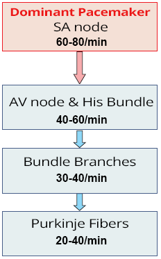
  • The conduction system spontaneously generates impulses
    • Impulses at the highest frequency are generated by the SA node (60-80/min.)
    • The SA node is designated as the dominant (primary) pacemaker
  • If the SA node fails to generate impulses due to malfunction
    • Lower centers of the conduction system are activated
    • which generate impulses at a lower frequency

  • Suppression of the conduction system means that
    • Impulses always spread through the conduction system from the site
      • that generates impulses at the highest frequency
    • The site with the highest frequency deactivates sites
      • that generate impulses at a lower frequency
      • This phenomenon is also referred to as overdrive suppression

Dominant pacemaker - SA node and overdrive suppression

ECG and Action Potential

Action potential duration and one electrical cardiac cycle

ECG (lead V6) and Action Potential

  • QRS complex represents ventricular depolarization
    • During the QRS complex, a depolarization wave spreads through the ventricles
  • ST segment is an isoelectric line
  • T wave represents ventricular repolarization
    • The electrical vector during repolarization has the same direction as during depolarization
    • However, repolarization takes longer, hence the T wave is wider than the QRS complex
  • QT interval corresponds to 1 electrical cardiac cycle:
    • Ventricular depolarization + repolarization
  • P wave represents atrial depolarization
    • The action potential of the P wave is not shown in the image
    • Atrial repolarization is hidden within the QRS complex

ECG and Cardiac Cycle

action potential, atrial diastole, atrial systole, ventricular diastole, ventricular systole, one cardiac cycle

ECG and Cardiac Cycle


Sources

  • ECG from Basics to Essentials Step by Step
  • litfl.com
  • ecgwaves.com
  • metealpaslan.com
  • medmastery.com
  • uptodate.com
  • ecgpedia.org
  • wikipedia.org
  • Strong Medicine
  • Understanding Pacemakers