ECGbook.com

Making Medical Education Free for All

ECGbook.com

Making Medical Education Free for All

Home /

Arrhythmias in Acute Myocardial Infarction

Arrhythmias and acute myocardial infarction

Coronary Blood Supply

Schematic representation of conduction system and blood supply, Ischemia and infarction
  • The heart is supplied by 3 main arteries:
    • Right Coronary Artery (RCA)
      • Posterior Interventricular Branch (RIP)
    • Left Coronary Artery (LCA):
      • Anterior Interventricular Branch (LAD)
      • Circumflex Branch (LCx)
  • The RIP determines coronary artery dominance:
    • RCA Dominance (RIP arises from RCA)
    • LCA Dominance (RIP arises from LCA)
    • Codominance (RIP arises from both RCA and LCA)
  • Each artery supplies a specific area through its branches:
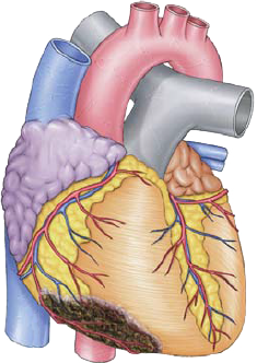
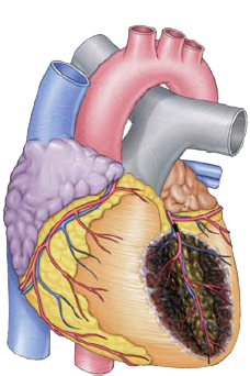
  • Infarction occurs in the area supplied by the occluded artery
  • Arrhythmias during infarction (ischemia) occur when
    • the infarction (ischemia) affects the conduction system
    • the infarction in the myocardium creates an active ectopic focus

  • Ischemia alters electrical properties
    • of both the conduction system and the myocardium
  • Infarction (necrosis) completely interrupts the conduction system

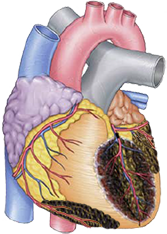
Infarction and Affected Artery


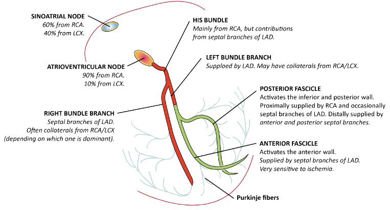
Blood Supply to the Conduction System
Conduction system blood supply: SA node, AV node, Bundle branches, PDA, LAD, LCx, culprit artery

Blood Supply to the Conduction System
  • SA Node
  • AV Node
    • Most commonly (90%) supplied by the RCA
    • Can also be supplied by the LCA (10%) through the LCx
  • Right Bundle Branch
  • His Bundle
    • Supplied by the RCA
    • Often also by collateral septal branches of LAD
  • Left Bundle Branch
    • Supplied by branches from LAD
    • Often also by collaterals from RCA and LCx
  • Left Posterior Fascicle
    • Proximal part supplied by RCA
    • Distal part supplied by septal branches of LAD
    • It is thick and relatively resistant to ischemia, left posterior block in ischemia is rare
  • Left Anterior Fascicle
    • Supplied by septal branches of LAD
    • It is thin, and very sensitive to ischemia, left anterior block in ischemia is common

Right Bundle Branch Block and Infarction

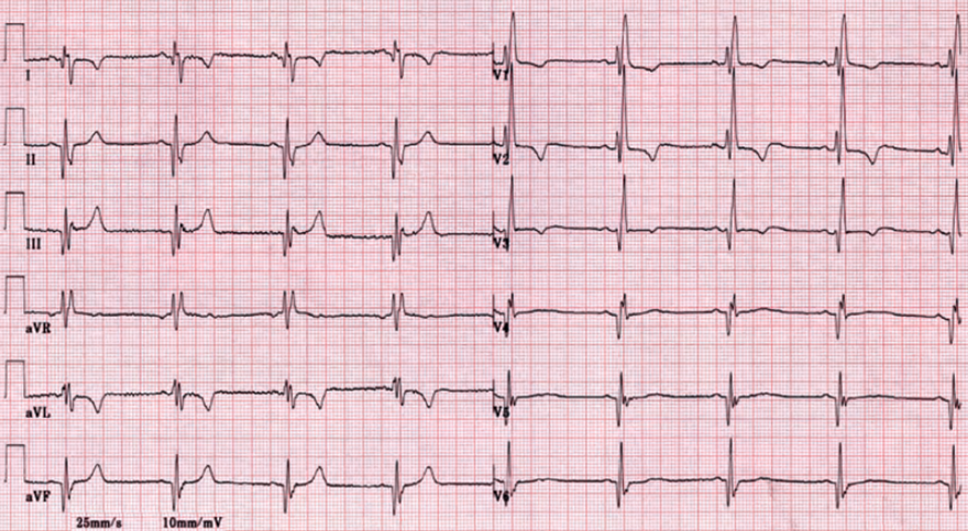
ECG RBBB and STEMI infarction Infarction due to proximal LAD occlusion (D1, S1) and RBBB

Left Bundle Branch Block and Infarction

ECG criteria V1 left bundle branch block (LBBB) and STEMI infarction


ECG criteria V6 left bundle branch block (LBBB) and STEMI infarction
  • LBBB primarily occurs due to a degenerative process in the conduction system
    • Prevalence of LBBB in the population:
      • At age 50, LBBB affects 1% of people
      • At age 80, LBBB affects 17% of people
  • Left Bundle Branch
    • Supplied by branches from LAD
    • Often also by collaterals from RCA and LCx
  • LBBB affects depolarization of the left ventricle
    • Infarction almost always occurs in the left ventricle
Proximal occlusion LAD (culprit artery) and left bundle branch block and STEMI infarction
  • LBBB can also occur with infarction due to a proximal occlusion of LAD
  • Up to 50% of patients with LBBB and infarction
    • may not have typical angina (chest pain)
  • LBBB creates on EKG:
    • ST elevations (V1-V3)
    • ST depressions (V5-V6, I, aVL)
  • Newly developed LBBB with symptoms of infarction
    • is considered an equivalent to STEMI
    • The issue is whether the LBBB is newly developed or old?
  • Infarction can occur:
    • Simultaneously with LBBB
    • In the setting of preexisting LBBB

  • Sgarbossa's EKG criteria
    • Used for diagnosing STEMI in the presence of LBBB

Arrhythmias in Inferior Wall Infarction

Arrhythmias in acute myocardial infarction, Inferior STEMI

Most Common Arrhythmias in Inferior STEMI

Arrhythmia Comment Prognosis
Sinus Bradycardia Is the most common
Occurs in 40% of inferior wall infarctions
Is temporary (max. 7 days)
SA Node Dysfunction Is very rare
Occurs in the subacute phase (hours - days)
Often permanent
AV Block I. Degree Is common
Occurs in the subacute phase (hours - days)
Is temporary (max. 7 days)
AV Block II. Degree (Type I) Is common
Occurs in the subacute phase (hours - days)
Is temporary (max. 7 days)
AV Block II. Degree (Type II) Is rare
However, often occurs in anterior infarction
Is temporary (max. 7 days)
AV Block III. Degree Is rare
Often develops progressively:
AV block I. -> II. -> III. degree
Is temporary (max. 7 days)

Arrhythmias in Anterior STEMI

Arrhythmias in acute anterior STEMI infarction

Most Common Arrhythmias in Anterior STEMI

Arrhythmia Comment Prognosis
AV Block II. Degree (Type II) Often progresses to AV Block III. Degree Often permanent
Bifascicular Block RBBB + LPH
  • Supplied by the same septal branches from RIA
  • Often progresses to AV Block III. Degree
Often permanent
AV Block III. Degree Often precedes
  • RBBB
  • Bifascicular Block
Has 80% mortality
Often permanent

Tachy and Brady Arrhythmias in Acute Myocardial Infarction


Reperfusion and Arrhythmias in Myocardial Infarction

STEMI infarction reperfusion arrhythmias, angioplasty, Resolution ST elevation, accelerated idioventricular rhythm
  • In STEMI infarction, ST elevations persist for 12-24 hours
  • Treatment of the infarction is recanalization (restoration of blood flow)
  • Decrease in ST elevations by 50% within 60-90 minutes after therapy
    • Is a positive marker of myocardial reperfusion
    • Improves patient prognosis
    • Q wave often disappears if it was present on the ECG
  • Decrease in ST elevations by 70% within 60-90 minutes after therapy
    • Is a marker of very good patient prognosis
    • Q wave often disappears if it was present on the ECG
  • Inverted T waves after reperfusion in the first hours of STEMI
    • Are a positive marker of successful reperfusion
  • Q wave in STEMI does not indicate irreversible damage to the myocardium
    • If a Q wave developed within 6 hours, it often disappears after successful reperfusion
  • 50% of patients have accelerated ventricular rhythm upon reperfusion
    • This is a benign arrhythmia that does not require treatment and resolves spontaneously

  • Markers of unsuccessful reperfusion and progression of occlusion include:


Old inferior, lateral, anterior infarction
ECG right bundle branch block, old infero-lateral infarction, old anterior infarction

Right Bundle Branch Block and Old Infarction


Sources

  • ECG from Basics to Essentials Step by Step
  • litfl.com
  • ecgwaves.com
  • metealpaslan.com
  • medmastery.com
  • uptodate.com
  • ecgpedia.org
  • wikipedia.org
  • Strong Medicine
  • Understanding Pacemakers


šípka späť

Arrhythmias in Acute Myocardial Infarction

Arrhythmias and acute myocardial infarction

Coronary Blood Supply

  • The heart is supplied by 3 main arteries:
    • Right Coronary Artery (RCA)
      • Posterior Interventricular Branch (RIP)
    • Left Coronary Artery (LCA):
      • Anterior Interventricular Branch (LAD)
      • Circumflex Branch (LCx)
  • The RIP determines coronary artery dominance:
    • RCA Dominance (RIP arises from RCA)
    • LCA Dominance (RIP arises from LCA)
    • Codominance (RIP arises from both RCA and LCA)
  • Each artery supplies a specific area through its branches:
  • Infarction occurs in the area supplied by the occluded artery
  • Arrhythmias during infarction (ischemia) occur when
    • the infarction (ischemia) affects the conduction system
    • the infarction in the myocardium creates an active ectopic focus

  • Ischemia alters electrical properties
    • of both the conduction system and the myocardium
  • Infarction (necrosis) completely interrupts the conduction system


Schematic representation of conduction system and blood supply, Ischemia and infarction

Infarction and Affected Artery


Blood Supply to the Conduction System

Conduction system blood supply: SA node, AV node, Bundle branches, PDA, LAD, LCx, culprit artery

Blood Supply to the Conduction System

  • SA Node
  • AV Node
    • Most commonly (90%) supplied by the RCA
    • Can also be supplied by the LCA (10%) through the LCx
  • Right Bundle Branch
  • His Bundle
    • Supplied by the RCA
    • Often also by collateral septal branches of LAD
  • Left Bundle Branch
    • Supplied by branches from LAD
    • Often also by collaterals from RCA and LCx
  • Left Posterior Fascicle
    • Proximal part supplied by RCA
    • Distal part supplied by septal branches of LAD
    • It is thick and relatively resistant to ischemia, left posterior block in ischemia is rare
  • Left Anterior Fascicle
    • Supplied by septal branches of LAD
    • It is thin, and very sensitive to ischemia, left anterior block in ischemia is common

Right Bundle Branch Block and Infarction

  • Prevalence of Right Bundle Branch Block (RBBB) in infarction is 7%
  • Right Bundle Branch
    • Supplied by septal branches from LAD
    • Often also by collaterals from RCA and LCx
  • RBBB in infarction usually occurs with left anterior hemiblock
    • Because they share the same blood supply via LAD
    • These blocks are collectively referred to as bifascicular block
  • RBBB is a disorder of depolarization of the thin right ventricle
    • Depolarization of the massive left ventricle remains intact
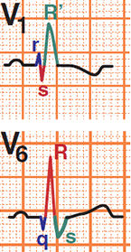
  • EKG and RBBB:
    • Wide QRS (≥0.12s)
    • rsR' (V1-V3) and deep S (V5-V6)
    • Slight ST depressions and negative T waves (V1-V3)

  • RBBB does not affect EKG criteria for diagnosing infarction
  • New RBBB in anterior STEMI indicates
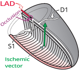
    • Proximal LAD occlusion (above S1 and D1 branches)
    • Right Bundle Branch is supplied by the S1 branch of LAD
  • RBBB can mask posterior STEMI
    • Because both conditions create ST depressions (V1-V3)
    • Posterior STEMI has ST elevations in posterior leads (V7-V9)
  • Pseudonormalization
    • RBBB shows ST depressions and negative T waves (V1-V3)
    • In right ventricle STEMI, leads (V1-V3) may normalize
    • Hyperacute STEMI causes ST elevations and peaked T waves
      • Thus, the ST segment and T waves normalize
      • This is known as "pseudonormalization"
ECG RBBB and STEMI infarction


Infarction due to proximal LAD occlusion (D1, S1) and RBBB

Left Bundle Branch Block and Infarction

  • LBBB primarily occurs due to a degenerative process in the conduction system
    • Prevalence of LBBB in the population:
      • At age 50, LBBB affects 1% of people
      • At age 80, LBBB affects 17% of people
  • Left Bundle Branch
    • Supplied by branches from LAD
    • Often also by collaterals from RCA and LCx
  • LBBB affects depolarization of the left ventricle
    • Infarction almost always occurs in the left ventricle
  • LBBB can also occur with infarction due to a proximal occlusion of LAD
  • Up to 50% of patients with LBBB and infarction
    • may not have typical angina (chest pain)

  • LBBB creates on EKG:
    • ST elevations (V1-V3)
    • ST depressions (V5-V6, I, aVL)
  • Newly developed LBBB with symptoms of infarction
    • is considered an equivalent to STEMI
    • The issue is whether the LBBB is newly developed or old?
  • Infarction can occur:
    • Simultaneously with LBBB
    • In the setting of preexisting LBBB

  • Sgarbossa's EKG criteria
    • Used for diagnosing STEMI in the presence of LBBB
ECG criteria V1 left bundle branch block (LBBB) and STEMI infarction

ECG criteria V6 left bundle branch block (LBBB) and STEMI infarction


Proximal occlusion LAD (culprit artery) and left bundle branch block and STEMI infarction

Arrhythmias in Inferior Wall Infarction


Arrhythmias in acute myocardial infarction, Inferior STEMI

Most Common Arrhythmias in Inferior STEMI

Arrhythmia Comment Prognosis
Sinus Bradycardia Is the most common
Occurs in 40% of inferior wall infarctions
Is temporary (max. 7 days)
SA Node Dysfunction Is very rare
Occurs in the subacute phase (hours - days)
Often permanent
AV Block I. Degree Is common
Occurs in the subacute phase (hours - days)
Is temporary (max. 7 days)
AV Block II. Degree (Type I) Is common
Occurs in the subacute phase (hours - days)
Is temporary (max. 7 days)
AV Block II. Degree (Type II) Is rare
However, often occurs in anterior infarction
Is temporary (max. 7 days)
AV Block III. Degree Is rare
Often develops progressively:
AV block I. -> II. -> III. degree
Is temporary (max. 7 days)

Arrhythmias in Anterior STEMI

  • Arrhythmias in anterior STEMI
    • Occur due to necrosis of the conduction system
    • Are permanent (do not resolve within 7 days)
  • Results in necrosis of the ventricular septum and His bundle
    • Escape rhythm activates distally from the His bundle
    • Therefore, QRS complexes are wide (>0.12s)

  • AV blocks in anterior STEMI often progress to
  • Anterior STEMI with AV block III. degree has an 80% mortality rate
Arrhythmias in acute anterior STEMI infarction

Most Common Arrhythmias in Anterior STEMI

Arrhythmia Comment Prognosis
AV Block II. Degree (Type II) Often progresses to AV Block III. Degree Often permanent
Bifascicular Block RBBB + LPH
  • Supplied by the same septal branches from RIA
  • Often progresses to AV Block III. Degree
Often permanent
AV Block III. Degree Often precedes
  • RBBB
  • Bifascicular Block
Has 80% mortality
Often permanent

Tachy and Brady Arrhythmias in Acute Myocardial Infarction


Reperfusion and Arrhythmias in Myocardial Infarction

  • In STEMI infarction, ST elevations persist for 12-24 hours
  • Treatment of the infarction is recanalization (restoration of blood flow)
  • Decrease in ST elevations by 50% within 60-90 minutes after therapy
    • Is a positive marker of myocardial reperfusion
    • Improves patient prognosis
    • Q wave often disappears if it was present on the ECG
  • Decrease in ST elevations by 70% within 60-90 minutes after therapy
    • Is a marker of very good patient prognosis
    • Q wave often disappears if it was present on the ECG
  • Inverted T waves after reperfusion in the first hours of STEMI
    • Are a positive marker of successful reperfusion
  • Q wave in STEMI does not indicate irreversible damage to the myocardium
    • If a Q wave developed within 6 hours, it often disappears after successful reperfusion
  • 50% of patients have accelerated ventricular rhythm upon reperfusion
    • This is a benign arrhythmia that does not require treatment and resolves spontaneously

  • Markers of unsuccessful reperfusion and progression of occlusion include:


STEMI infarction reperfusion arrhythmias, angioplasty, Resolution ST elevation, accelerated idioventricular rhythm


ECG right bundle branch block, old infero-lateral infarction, old anterior infarction

Right Bundle Branch Block and Old Infarction

Old inferior, lateral, anterior infarction


Sources

  • ECG from Basics to Essentials Step by Step
  • litfl.com
  • ecgwaves.com
  • metealpaslan.com
  • medmastery.com
  • uptodate.com
  • ecgpedia.org
  • wikipedia.org
  • Strong Medicine
  • Understanding Pacemakers