|
|
ECGbook.com Making Medical Education Free for All |
Upload ECG for Interpretation |
|
|
ECGbook.com Making Medical Education Free for All |
Upload ECG for Interpretation |
|
|
ECGbook.com Making Medical Education Free for All |
Home /
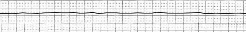
Asystole, Sudden cardiac arrest (SCA), Sudden cardiac death (SCD), Pulseless electrical activity (PEA)
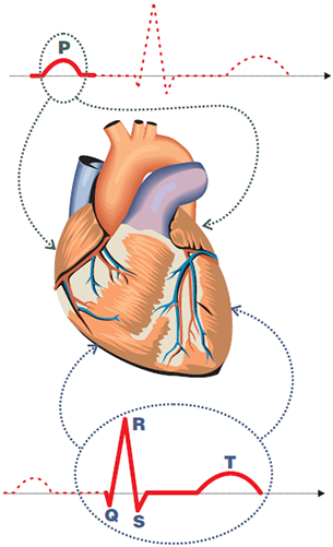

ECG and Cardiac Cycle
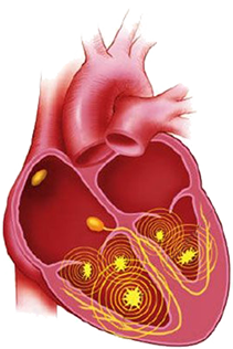
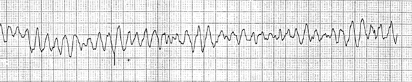
Ventricular Fibrillation

Ventricular Fibrillation and Asystole
Sources
Home /
Asystole, Sudden cardiac arrest (SCA), Sudden cardiac death (SCD), Pulseless electrical activity (PEA)
ECG and Electrical Impulse
|
|
ECG and Cardiac Cycle
|
|
Ventricular Fibrillation
|
Ventricular Fibrillation and Asystole
Sources