ECGbook.com

Making Medical Education Free for All

ECGbook.com

Making Medical Education Free for All

Home /

Athletes and ECG

ECG interpretation in athletes

Athlete's Heart

ECG Athletic heart

Sudden Cardiac Death


Normal ECG in Athletes

  • The table shows ECG findings
    • that are considered a variant of normal in athletes
  • An asymptomatic athlete with these ECG findings
    • does not require further cardiac examinations

Normal ECG in athletes:
Sinus bradycardia ≤ 30/min.
Sinus respiratory arrhythmia
Ectopic atrial rhythm
Junctional rhythm
First-degree AV block
Second-degree AV block (Wenckebach)
Incomplete RBBB
  • Occurs in approximately 30-50% of athletes
Only voltage criteria for left ventricular hypertrophy
  • No other criteria: axis deviation, P mitrale...
Benign early repolarization
Convex ST elevations and negative T waves (V1-V4)
  • African/black athletes


Abnormal ECG in Athletes


Abnormal ECG in Athletes:
Negative T waves
  • >1mm in at least 2 leads: (V2-V6, II, aVF, I, aVL)
ST depression (≥0.5mm in at least 2 leads)
Pathological Q waves
  • >3mm, or >40ms in at least 2 leads (excluding III, aVR)
Complete LBBB
Prolonged ventricular conduction (QRS≥0.14s)
Left axis deviation
P mitrale
Right ventricular hypertrophy
Pre-excitation
Prolonged QT interval
Shortened QT interval
Brugada syndrome
Sinus bradycardia (<30/min.)
Sinus pause (≥3sec.)
Supraventricular tachycardia (excluding sinus tachycardia)
Ventricular extrasystole (≥2 VES in 10s, couplet, triplet)
Ventricular tachycardia


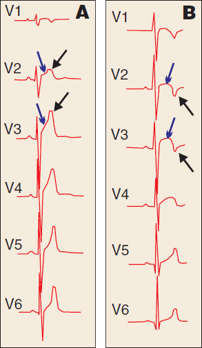
ECG in two healthy athletes, early repolarization, concave and convex ST segment elevation, negative T wave, positive T wave

Normal ECG in Athletes

  • These are ECG recordings from 2 healthy athletes
  • A: Benign early repolarization
    • Concave ST elevations and positive T waves
  • B: Benign early repolarization
    • Convex ST elevations and negative T waves


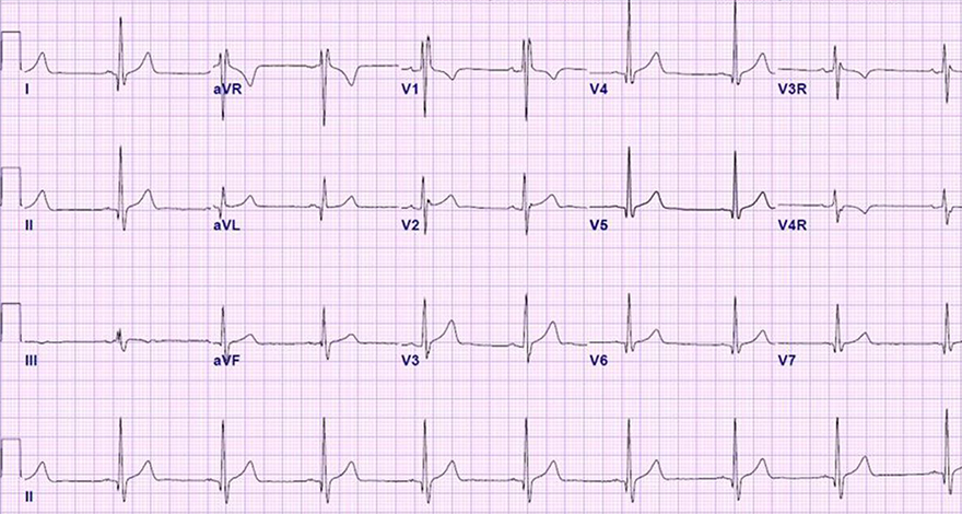
ECG healthy athletes, incomplete right bundle branch block (RBBB)

Normal ECG in an Athlete


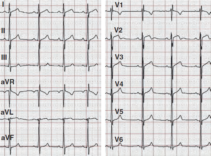
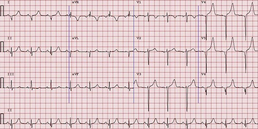
ECG athlete, sport man, sinus bradycardia, early repolarisation, peaked T waves, voltage criteria for left ventricular hypertrophy

Normal ECG in an Athlete


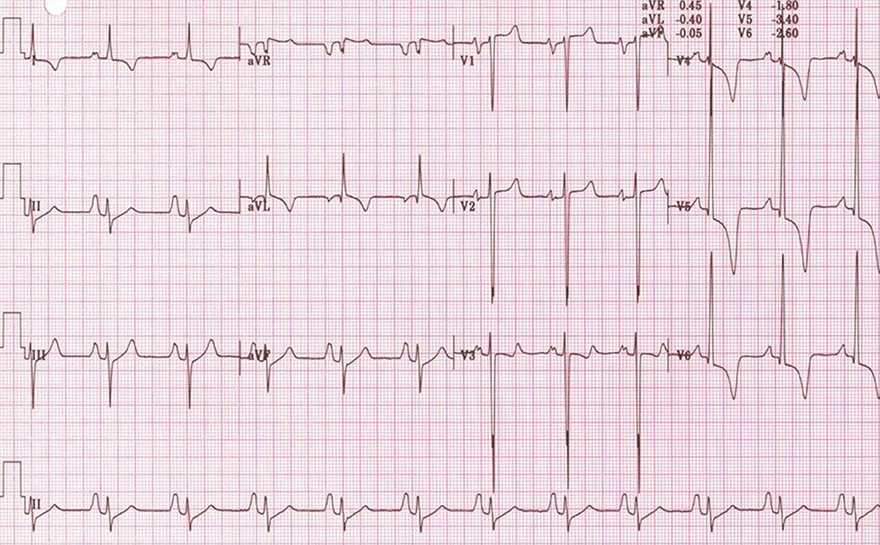
ECG athlete hypertrophic cardiomyopathy, QRS voltage criteria for LVH, deep TWI and ST segment depression, left atrial and right atrial enlargement, left axis deviation

Abnormal ECG in an Athlete


ECG athlete hypertrophic cardiomyopathy, QRS voltage criteria for LVH, deep TWI and ST segment depression, left atrial and right atrial enlargement, left axis deviation

Normal ECG in an Athlete

  • Incomplete right bundle branch block
    • rSR (V1)
    • QRS < 120ms
  • This is a normal ECG finding in an athlete


ECG black athlete, elevation and convex ST segment elevation followed by T wave inversion in V1-V4, voltage left ventricular hypertrophy

Normal ECG in an Athlete


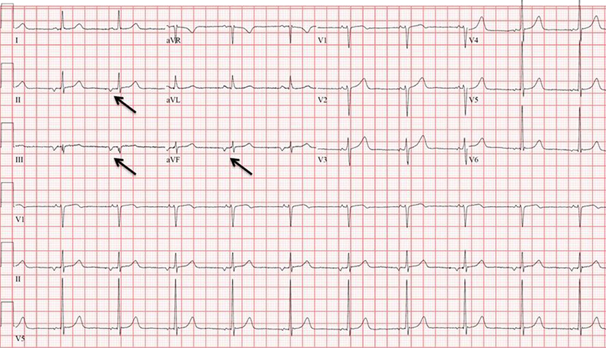
ECG athlete, junctional escape rhythm

Normal ECG in an Athlete

  • This is a 28-year-old athlete
  • Junctional rhythm (arrows)
  • This is a normal finding in athletes


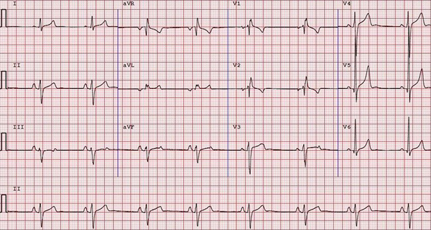
ECG athlete,  ectopic atrial rhythm, Negative P wave in inferior leads (II, III, aVF)

Normal ECG in an Athlete


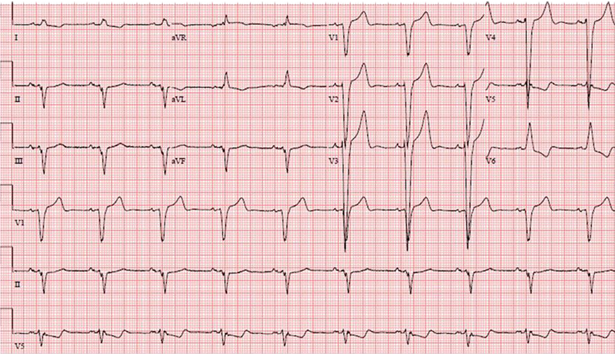
Abnormal ECG athlete complete right bundle branch block, left axis deviation,  right atrial enlargement - P pulmonale

Abnormal ECG in an Athlete


Abnormal ECG athlete deep pathological Q wave in V4-V6, I, aVL, hypertrophic cardiomyopathy

Abnormal ECG in an Athlete


Abnormal ECG athlete left bundle branch block (LBBB)

Abnormal ECG in an Athlete

  • This is a 26-year-old athlete
  • Left bundle branch block
    • Is always considered an abnormal ECG finding in athletes
  • This is an abnormal ECG finding
    • The athlete must undergo further heart-focused examinations


Abnormal ECG athlete, multiple premature ventricular complexes, PVC LBBB pattern

Abnormal ECG in an Athlete


Sources

  • ECG from Basics to Essentials Step by Step
  • litfl.com
  • ecgwaves.com
  • metealpaslan.com
  • medmastery.com
  • uptodate.com
  • ecgpedia.org
  • wikipedia.org
  • Strong Medicine
  • Understanding Pacemakers


šípka späť

Athletes and ECG

ECG interpretation in athletes

Athlete's Heart

ECG Athletic heart

Sudden Cardiac Death


Normal ECG in Athletes

  • The table shows ECG findings
    • that are considered a variant of normal in athletes
  • An asymptomatic athlete with these ECG findings
    • does not require further cardiac examinations

Normal ECG in athletes:
Sinus bradycardia ≤ 30/min.
Sinus respiratory arrhythmia
Ectopic atrial rhythm
Junctional rhythm
First-degree AV block
Second-degree AV block (Wenckebach)
Incomplete RBBB
  • Occurs in approximately 30-50% of athletes
Only voltage criteria for left ventricular hypertrophy
  • No other criteria: axis deviation, P mitrale...
Benign early repolarization
Convex ST elevations and negative T waves (V1-V4)
  • African/black athletes


Abnormal ECG in Athletes


Abnormal ECG in Athletes:
Negative T waves
  • >1mm in at least 2 leads: (V2-V6, II, aVF, I, aVL)
ST depression (≥0.5mm in at least 2 leads)
Pathological Q waves
  • >3mm, or >40ms in at least 2 leads (excluding III, aVR)
Complete LBBB
Prolonged ventricular conduction (QRS≥0.14s)
Left axis deviation
P mitrale
Right ventricular hypertrophy
Pre-excitation
Prolonged QT interval
Shortened QT interval
Brugada syndrome
Sinus bradycardia (<30/min.)
Sinus pause (≥3sec.)
Supraventricular tachycardia (excluding sinus tachycardia)
Ventricular extrasystole (≥2 VES in 10s, couplet, triplet)
Ventricular tachycardia


ECG in two healthy athletes, early repolarization, concave and convex ST segment elevation, negative T wave, positive T wave

Normal ECG in Athletes

  • These are ECG recordings from 2 healthy athletes
  • A: Benign early repolarization
    • Concave ST elevations and positive T waves
  • B: Benign early repolarization
    • Convex ST elevations and negative T waves


ECG healthy athletes, incomplete right bundle branch block (RBBB)

Normal ECG in an Athlete


ECG athlete, sport man, sinus bradycardia, early repolarisation, peaked T waves, voltage criteria for left ventricular hypertrophy

Normal ECG in an Athlete


ECG athlete hypertrophic cardiomyopathy, QRS voltage criteria for LVH, deep TWI and ST segment depression, left atrial and right atrial enlargement, left axis deviation

Abnormal ECG in an Athlete


ECG athlete hypertrophic cardiomyopathy, QRS voltage criteria for LVH, deep TWI and ST segment depression, left atrial and right atrial enlargement, left axis deviation

Normal ECG in an Athlete

  • Incomplete right bundle branch block
    • rSR (V1)
    • QRS < 120ms
  • This is a normal ECG finding in an athlete


ECG black athlete, elevation and convex ST segment elevation followed by T wave inversion in V1-V4, voltage left ventricular hypertrophy

Normal ECG in an Athlete


ECG athlete, junctional escape rhythm

Normal ECG in an Athlete

  • This is a 28-year-old athlete
  • Junctional rhythm (arrows)
  • This is a normal finding in athletes


ECG athlete,  ectopic atrial rhythm, Negative P wave in inferior leads (II, III, aVF)

Normal ECG in an Athlete


Abnormal ECG athlete complete right bundle branch block, left axis deviation,  right atrial enlargement - P pulmonale

Abnormal ECG in an Athlete


Abnormal ECG athlete deep pathological Q wave in V4-V6, I, aVL, hypertrophic cardiomyopathy

Abnormal ECG in an Athlete


Abnormal ECG athlete left bundle branch block (LBBB)

Abnormal ECG in an Athlete

  • This is a 26-year-old athlete
  • Left bundle branch block
    • Is always considered an abnormal ECG finding in athletes
  • This is an abnormal ECG finding
    • The athlete must undergo further heart-focused examinations


Abnormal ECG athlete, multiple premature ventricular complexes, PVC LBBB pattern

Abnormal ECG in an Athlete


Sources

  • ECG from Basics to Essentials Step by Step
  • litfl.com
  • ecgwaves.com
  • metealpaslan.com
  • medmastery.com
  • uptodate.com
  • ecgpedia.org
  • wikipedia.org
  • Strong Medicine
  • Understanding Pacemakers