ECGbook.com

Making Medical Education Free for All

ECGbook.com

Making Medical Education Free for All

Home /

Atrial Fibrillation

Atrial Fibrillation (AF or A-fib)

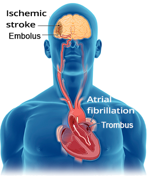
Atrial Fibrillation and Stroke

    Thromboembolic ischemic stroke, atrial fibrillation, left atrial appendage with thrombus formation
  • Atrial Fibrillation (AF) is the most common arrhythmia, with an occurrence of:
    • approximately 50,000 people in Slovakia
    • approximately 4.5 million people in the European Union
  • Every year, 400 people in Slovakia suffer a stroke
    • 80 strokes annually in Slovakia are caused by AF

  • In AF, the atria "quiver" with a frequency of 350-600/min.
    • Blood stagnates in the atria and forms a thrombus
      • Most commonly in the left atrial appendage
    • The thrombus may detach (detached thrombus = embolus)
      • and travel to the brain (where it narrows a vessel)
    • Thus, a patient with AF can suffer from a thromboembolic stroke
    • Atrial flutter has the same mechanism
  • The risk of thrombus formation is the same regardless of the duration of AF
    • It does not matter if the patient has had AF for 7 days or 3 months
    • Patients with silent (asymptomatic) AF are at higher risk
      • which the patients do not feel
  • Vicious Circle
    • AF causes both organic and electrical remodeling of the atria
    • thus creating a substrate for AF

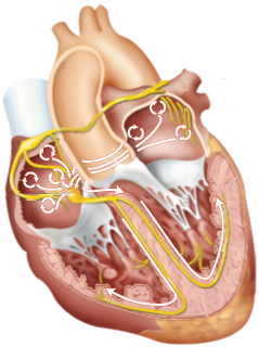
Atria and Atrial Fibrillation

Heart with atrial fibrillation

Multifocal Atrial Activity in Atrial Fibrillation

  • Atrial fibrillation requires:
    • Substrate (electrically or structurally altered atria) to sustain abnormal electrical activity
    • Trigger (most commonly atrial ectopy)
  • Atrial fibrillation is multifocal activity in the atria
    • The atria have a number of foci that independently generate impulses
      • with a frequency of 350-600/min., the impulses outpace the SA node and "turn it off" (overdrive suppression)

  • The mechanism of multifocal atrial activity during AF is uncertain; there are 3 theories:
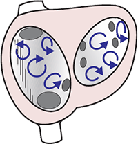
    • Numerous micro re-entries
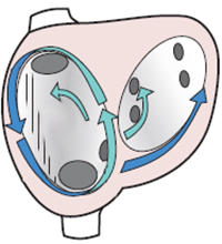
    • Mother focus and activation waves
    • Mother re-entry and activation waves


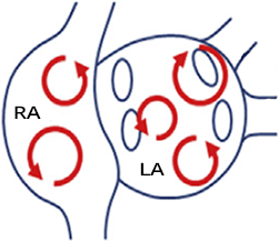
Atrial fibrillation, multiple wavelet mechanism, multiple micro re-entry

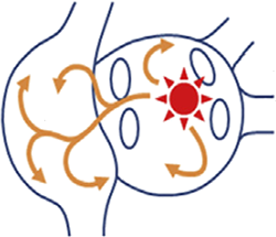
Atrial fibrillation, focal activity, increased automaticity, micro re-entry

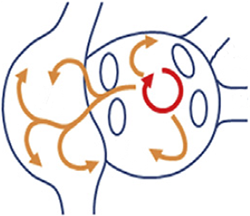
Focal Focus and Activation Waves

  • In the atrium, there is a focal focus with increased automaticity
  • The focus generates numerous activation waves, each of which activates the atria
    • The activation waves can create micro re-entries
  • The most common trigger is again atrial ectopy
  • The atria have a frequency of 350-600/min.


Atrial fibrillation, mother rotors re-entry, multiple wavelet mechanism

Maternal Re-entry and Activation Waves


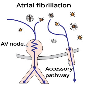
Protective Mechanism of the AV Node

Atrial fibrillation with accessory pathway and WPW syndrome, decremental and non-decremental conduction delay
  • Impulses to the ventricles do not pass through the AV node at 350-600/min.
  • In WPW syndrome
    • An accessory pathway (connecting atria and ventricles) is present
      • The pathway does not have a protective mechanism like the AV node
    • A patient with WPW and atrial fibrillation
      • must not receive medications to slow down the AV node
        • Impulses would pass to the ventricles only through the pathway
        • At a frequency of 350-600/min.
        • Resulting in ventricular fibrillation

ECG and Atrial Fibrillation


ECG coarse atrial fibrillation, irregularly irregular rhythm, no P waves, narrow QRS

Atrial Fibrillation


Frequency Calculation

  • For irregular heart rhythms, the 6-second method is used to calculate the frequency
    • This is the average frequency of QRS complexes over 6 seconds (30 squares)
    • Frequency = number of QRS complexes in 6s x 10
Atrial fibrillation, irregular rhythm, 6 seconds method, heart rate

Atrial Fibrillation and Frequency 130/min.

  • Atrial fibrillation
  • Number of QRS complexes in 6s (30 squares) is 13
  • 13 x 10 = 130/min.

Ashman Phenomenon

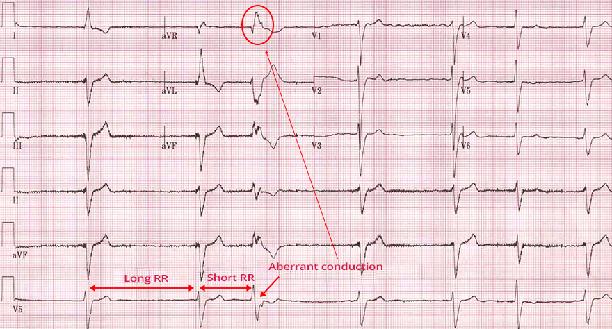
Atrial fibrillation Ashman phenomenon, long refractory period, RR interval, aberrantly conducted beat RBBB morphology


ECG atrial fibrillation, ashman phenomenon, long RR, short RR, aberration RBBB morphology

Ashman Phenomenon and Atrial Fibrillation

  • On the ECG, atrial fibrillation is seen in lead V1 with a calibration of 10mm
  • Initially, the RR intervals are relatively consistent
  • Then a long-short RR interval pattern follows
    • The long RR interval extends the refractory period
    • The short RR interval ends with aberrant conduction showing the appearance of a Right Tawar Bundle Block (RTB)
      • The short RR interval impulse passes through the right bundle during the refractory period and gets blocked
  • Ashman phenomenon is most commonly observed in atrial fibrillation
    • When RR intervals change irregularly

Concealed Conduction

ECG, laddergram concealed conduction and atrial fibrillation

Classification of Atrial Fibrillation by Frequency

  • The AV node most commonly transmits impulses to the ventricles at a rate of 110-160/min.
  • Based on the ventricular frequency (QRS complexes), atrial fibrillation can be classified into:

Differential Diagnosis of Atrial Fibrillation


atrial fibrillation, multifocal re-entry
ECG atrial fibrillation, irregular rhythm

Atrial Fibrillation

  • In the atria, there are many micro re-entry circuits
    • They generate impulses independently with a frequency of 350-600/min
  • The heart rhythm is irregularly irregular (the RR intervals are of varying lengths)
  • Fibrillatory f-waves (deformed baseline, no P waves)


atrial flutter alternating 2:1, 4:1 conduction ratios, macro re-entry
ECG atrial flutter, conduction ratio 2:1, 4:1

Atrial Flutter with Variable Conduction (2:1 and 4:1)


Multifocal atrial tachycardia mechanism
ECG multifocal atrial tachycardia, enhanced automaticity

Multifocal Atrial Tachycardia

  • Multifocal Atrial Tachycardia
  • There are 3 ectopic foci in the atria generating impulses independently
  • On the ECG, there are 3 different P waves
    • The ECG shows 3 P waves of different shapes (each focus generates its own P wave)
  • The heart rhythm is irregularly irregular (similar to atrial fibrillation)
    • Because the ectopic foci generate impulses (P waves) independently of each other
    • However, each P wave is followed by a QRS complex

Classification of Atrial Fibrillation


  • In addition to the duration, atrial fibrillation (AF) can also be classified according to the following criteria:

  • Newly Diagnosed Atrial Fibrillation
    • AF is recorded once on an ECG
    • Sometimes atrial fibrillation is diagnosed incidentally during a preventive examination
      • Silent atrial fibrillation is asymptomatic (patients do not feel it)
  • Silent Atrial Fibrillation
    • It is asymptomatic atrial fibrillation
    • The patient does not feel it; it is very insidious because it is often untreated
  • Isolated (Lone) Atrial Fibrillation
    • Accounts for 20% of all AF cases
    • Sometimes referred to as idiopathic atrial fibrillation (AF of unknown origin)
    • Occurs in structurally normal atria from an unknown cause?
  • Non-Valvular Atrial Fibrillation
    • AF occurs in the heart without:
      • Valvular heart disease
      • Artificial valves
  • Valvular Atrial Fibrillation
    • AF occurs in the heart with valvular heart disease or with an artificial valve


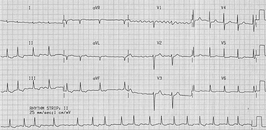
ECG coarse atrial fibrillation, irregular ventricular response

Atrial Fibrillation


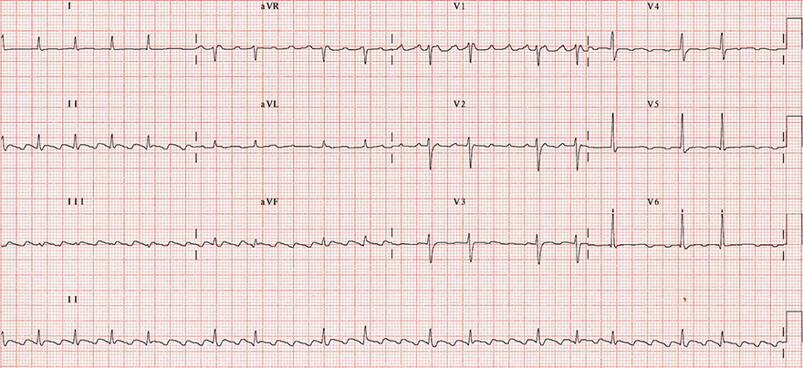
ECG typical atrial flutter, ratio 2:1, 4:1, variable AV block

Atrial Flutter (with Variable Conduction 2:1 and 4:1)

  • The heart rhythm is regularly irregular, with 2 RR intervals alternating
    • One RR interval has a 2:1 conduction to the ventricles
    • The other RR interval has a 4:1 conduction to the ventricles
  • This is a Typical Isthmus-dependent Flutter (impulse circulates counterclockwise in the right atrium)
    • Inverted flutter waves in the lower leads (II, III, aVF) with a rate of 300/min.
    • Positive flutter waves in V1 (resembling P waves)
  • The ventricular conduction alternates between 2:1 and 4:1 (note the continuous lead II)
  • The RR interval with 4:1 block is exactly twice the RR interval with 2:1 block


ECG coarse atrial fibrillation, irregular narrow-complex, coarse fibrillatory waves

Atrial Fibrillation


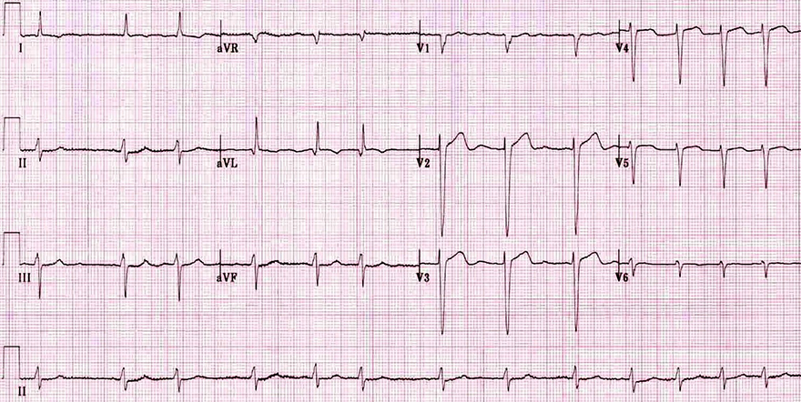
ECG fine atrial fibrillation, fine fibrillatory waves seen in V1, irregular ventricular response

Atrial Fibrillation


ECG multifocal atrial tachycardia, irreguler rhythm, constant PR interval

Multifocal Atrial Tachycardia


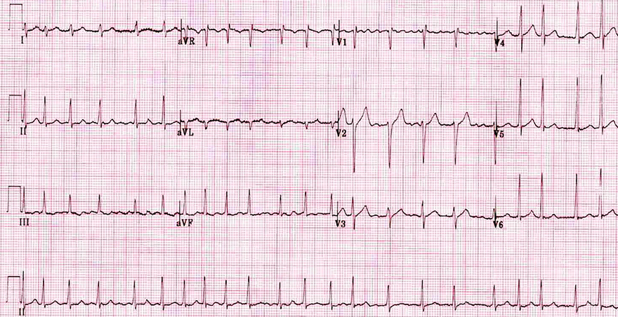
ECG coarse atrial fibrillation, rapid ventricular response, irregular narrow-complex tachycardia

Atrial Fibrillation


ECG fine atrial fibrillation, slow ventricular rate, irregular heart rate, fibrillatory waves

Atrial Fibrillation


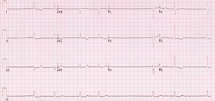
ECG ashman phenomenon, atrial fibrillation, aberrant conduction, RBBB

Atrial Fibrillation and Ashman’s Phenomenon

  • The heart rhythm is irregularly irregular
  • This is atrial fibrillation with a slow ventricular response
  • This is fine atrial fibrillation
    • Amplitude of fibrillation waves in V1 is <0.5mm
  • Ashman’s phenomenon (continuous V5 lead)
    • The 3rd QRS complex is wider and has a negative T wave
    • The AV node conducted an atrial impulse to the ventricles at a time when the right bundle branch was in a refractory period


ECG atrial fibrillation, ashman phenomenon, aberrant concuction, narrow qrs

Atrial Fibrillation and Ashman’s Phenomenon


Sources

  • ECG from Basics to Essentials Step by Step
  • litfl.com
  • ecgwaves.com
  • metealpaslan.com
  • medmastery.com
  • uptodate.com
  • ecgpedia.org
  • wikipedia.org
  • Strong Medicine
  • Understanding Pacemakers


šípka späť

Atrial Fibrillation

Atrial Fibrillation (AF or A-fib)

Atrial Fibrillation and Stroke

  • Atrial Fibrillation (AF) is the most common arrhythmia, with an occurrence of:
    • approximately 50,000 people in Slovakia
    • approximately 4.5 million people in the European Union
  • Every year, 400 people in Slovakia suffer a stroke
    • 80 strokes annually in Slovakia are caused by AF

  • In AF, the atria "quiver" with a frequency of 350-600/min.
    • Blood stagnates in the atria and forms a thrombus
      • Most commonly in the left atrial appendage
    • The thrombus may detach (detached thrombus = embolus)
      • and travel to the brain (where it narrows a vessel)
    • Thus, a patient with AF can suffer from a thromboembolic stroke
    • Atrial flutter has the same mechanism
  • The risk of thrombus formation is the same regardless of the duration of AF
    • It does not matter if the patient has had AF for 7 days or 3 months
    • Patients with silent (asymptomatic) AF are at higher risk
      • which the patients do not feel
  • Vicious Circle
    • AF causes both organic and electrical remodeling of the atria
    • thus creating a substrate for AF
Thromboembolic ischemic stroke, atrial fibrillation, left atrial appendage with thrombus formation

Atria and Atrial Fibrillation


Heart with atrial fibrillation

Multifocal Atrial Activity in Atrial Fibrillation

  • Atrial fibrillation requires:
    • Substrate (electrically or structurally altered atria) to sustain abnormal electrical activity
    • Trigger (most commonly atrial ectopy)
  • Atrial fibrillation is multifocal activity in the atria
    • The atria have a number of foci that independently generate impulses
      • with a frequency of 350-600/min., the impulses outpace the SA node and "turn it off" (overdrive suppression)

  • The mechanism of multifocal atrial activity during AF is uncertain; there are 3 theories:
    • Numerous micro re-entries
    • Mother focus and activation waves
    • Mother re-entry and activation waves


Atrial fibrillation, multiple wavelet mechanism, multiple micro re-entry

Numerous Micro Re-entries

  • In the atria, there are numerous micro re-entries
    • They independently generate impulses with a frequency of 350-600/min.
      • In each re-entry, the impulse circulates around
    • There are at least 10 micro re-entry circuits
  • Micro re-entry circuits
    • Are most often triggered by atrial ectopy
    • Require a substrate (remodeled atria) to be maintained
  • If there are 3 micro re-entries, multifocal atrial tachycardia will develop


Atrial fibrillation, focal activity, increased automaticity, micro re-entry

Focal Focus and Activation Waves

  • In the atrium, there is a focal focus with increased automaticity
  • The focus generates numerous activation waves, each of which activates the atria
    • The activation waves can create micro re-entries
  • The most common trigger is again atrial ectopy
  • The atria have a frequency of 350-600/min.


Atrial fibrillation, mother rotors re-entry, multiple wavelet mechanism

Maternal Re-entry and Activation Waves

  • The mechanism is the same as above, but in the atria there is a maternal re-entry
  • From the maternal re-entry, numerous activation waves emerge, each activating the atria
    • The activation waves can again create micro re-entries
  • The atria have a frequency of 350-600/min.
  • One micro re-entry creates intra-atrial re-entry tachycardia

Protective Mechanism of the AV Node

  • Impulses to the ventricles do not pass through the AV node at 350-600/min.
  • In WPW syndrome
    • An accessory pathway (connecting atria and ventricles) is present
      • The pathway does not have a protective mechanism like the AV node
    • A patient with WPW and atrial fibrillation
      • must not receive medications to slow down the AV node
        • Impulses would pass to the ventricles only through the pathway
        • At a frequency of 350-600/min.
        • Resulting in ventricular fibrillation

Atrial fibrillation with accessory pathway and WPW syndrome, decremental and non-decremental conduction delay

ECG and Atrial Fibrillation


ECG coarse atrial fibrillation, irregularly irregular rhythm, no P waves, narrow QRS

Atrial Fibrillation


Frequency Calculation

  • For irregular heart rhythms, the 6-second method is used to calculate the frequency
    • This is the average frequency of QRS complexes over 6 seconds (30 squares)
    • Frequency = number of QRS complexes in 6s x 10
Atrial fibrillation, irregular rhythm, 6 seconds method, heart rate

Atrial Fibrillation and Frequency 130/min.

  • Atrial fibrillation
  • Number of QRS complexes in 6s (30 squares) is 13
  • 13 x 10 = 130/min.

Ashman Phenomenon

Atrial fibrillation Ashman phenomenon, long refractory period, RR interval, aberrantly conducted beat RBBB morphology


ECG atrial fibrillation, ashman phenomenon, long RR, short RR, aberration RBBB morphology

Ashman Phenomenon and Atrial Fibrillation

  • On the ECG, atrial fibrillation is seen in lead V1 with a calibration of 10mm
  • Initially, the RR intervals are relatively consistent
  • Then a long-short RR interval pattern follows
    • The long RR interval extends the refractory period
    • The short RR interval ends with aberrant conduction showing the appearance of a Right Tawar Bundle Block (RTB)
      • The short RR interval impulse passes through the right bundle during the refractory period and gets blocked
  • Ashman phenomenon is most commonly observed in atrial fibrillation
    • When RR intervals change irregularly

Concealed Conduction

ECG, laddergram concealed conduction and atrial fibrillation

Classification of Atrial Fibrillation by Frequency

  • The AV node most commonly transmits impulses to the ventricles at a rate of 110-160/min.
  • Based on the ventricular frequency (QRS complexes), atrial fibrillation can be classified into:

Differential Diagnosis of Atrial Fibrillation


atrial fibrillation, multifocal re-entry ECG atrial fibrillation, irregular rhythm

Atrial Fibrillation

  • In the atria, there are many micro re-entry circuits
    • They generate impulses independently with a frequency of 350-600/min
  • The heart rhythm is irregularly irregular (the RR intervals are of varying lengths)
  • Fibrillatory f-waves (deformed baseline, no P waves)


atrial flutter alternating 2:1, 4:1 conduction ratios, macro re-entry ECG atrial flutter, conduction ratio 2:1, 4:1

Atrial Flutter with Variable Conduction (2:1 and 4:1)
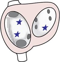
  • Atrial Flutter
  • There is one macro re-entry circuit around the entire right atrium
    • The impulse circles in the macro re-entry with a frequency of 300/min
  • Instead of P waves, the ECG shows Flutter (F) waves (sawtooth pattern) with a frequency of 300/min
  • The heart rhythm is regularly irregular
    • One RR interval has a 2:1 conduction ratio to the ventricles
    • The next RR interval has a 4:1 conduction ratio to the ventricles


Multifocal atrial tachycardia mechanism ECG multifocal atrial tachycardia, enhanced automaticity

Multifocal Atrial Tachycardia

  • Multifocal Atrial Tachycardia
  • There are 3 ectopic foci in the atria generating impulses independently
  • On the ECG, there are 3 different P waves
    • The ECG shows 3 P waves of different shapes (each focus generates its own P wave)
  • The heart rhythm is irregularly irregular (similar to atrial fibrillation)
    • Because the ectopic foci generate impulses (P waves) independently of each other
    • However, each P wave is followed by a QRS complex

Classification of Atrial Fibrillation


  • In addition to the duration, atrial fibrillation (AF) can also be classified according to the following criteria:

  • Newly Diagnosed Atrial Fibrillation
    • AF is recorded once on an ECG
    • Sometimes atrial fibrillation is diagnosed incidentally during a preventive examination
      • Silent atrial fibrillation is asymptomatic (patients do not feel it)
  • Silent Atrial Fibrillation
    • It is asymptomatic atrial fibrillation
    • The patient does not feel it; it is very insidious because it is often untreated
  • Isolated (Lone) Atrial Fibrillation
    • Accounts for 20% of all AF cases
    • Sometimes referred to as idiopathic atrial fibrillation (AF of unknown origin)
    • Occurs in structurally normal atria from an unknown cause?
  • Non-Valvular Atrial Fibrillation
    • AF occurs in the heart without:
      • Valvular heart disease
      • Artificial valves
  • Valvular Atrial Fibrillation
    • AF occurs in the heart with valvular heart disease or with an artificial valve


ECG coarse atrial fibrillation, irregular ventricular response

Atrial Fibrillation


ECG typical atrial flutter, ratio 2:1, 4:1, variable AV block

Atrial Flutter (with Variable Conduction 2:1 and 4:1)
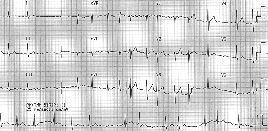
  • The heart rhythm is regularly irregular, with 2 RR intervals alternating
    • One RR interval has a 2:1 conduction to the ventricles
    • The other RR interval has a 4:1 conduction to the ventricles
  • This is a Typical Isthmus-dependent Flutter (impulse circulates counterclockwise in the right atrium)
    • Inverted flutter waves in the lower leads (II, III, aVF) with a rate of 300/min.
    • Positive flutter waves in V1 (resembling P waves)
  • The ventricular conduction alternates between 2:1 and 4:1 (note the continuous lead II)
  • The RR interval with 4:1 block is exactly twice the RR interval with 2:1 block


ECG coarse atrial fibrillation, irregular narrow-complex, coarse fibrillatory waves

Atrial Fibrillation


ECG fine atrial fibrillation, fine fibrillatory waves seen in V1, irregular ventricular response

Atrial Fibrillation


ECG multifocal atrial tachycardia, irreguler rhythm, constant PR interval

Multifocal Atrial Tachycardia


ECG coarse atrial fibrillation, rapid ventricular response, irregular narrow-complex tachycardia

Atrial Fibrillation


ECG fine atrial fibrillation, slow ventricular rate, irregular heart rate, fibrillatory waves

Atrial Fibrillation


ECG ashman phenomenon, atrial fibrillation, aberrant conduction, RBBB

Atrial Fibrillation and Ashman’s Phenomenon
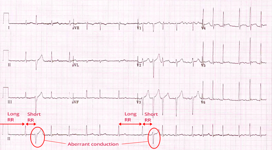
  • The heart rhythm is irregularly irregular
  • This is atrial fibrillation with a slow ventricular response
  • This is fine atrial fibrillation
    • Amplitude of fibrillation waves in V1 is <0.5mm
  • Ashman’s phenomenon (continuous V5 lead)
    • The 3rd QRS complex is wider and has a negative T wave
    • The AV node conducted an atrial impulse to the ventricles at a time when the right bundle branch was in a refractory period


ECG atrial fibrillation, ashman phenomenon, aberrant concuction, narrow qrs

Atrial Fibrillation and Ashman’s Phenomenon


Sources

  • ECG from Basics to Essentials Step by Step
  • litfl.com
  • ecgwaves.com
  • metealpaslan.com
  • medmastery.com
  • uptodate.com
  • ecgpedia.org
  • wikipedia.org
  • Strong Medicine
  • Understanding Pacemakers