|
|
ECGbook.com Making Medical Education Free for All |
Upload ECG for Interpretation |
|
|
ECGbook.com Making Medical Education Free for All |
Upload ECG for Interpretation |
|
|
ECGbook.com Making Medical Education Free for All |
Home /
Atrial myocardial infarction (ischemia), Ta wave elevation and depression, Atrial Ischemia, Atrial Injury

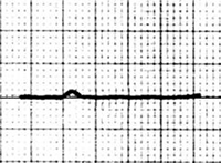


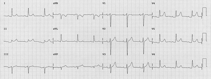
PQ Depression (1mm)
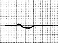
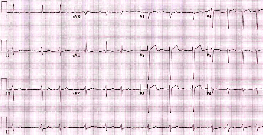
PQ Depression (2mm)

Atrial Infarction
Pericarditis

Right Atrial Infarction and Inferior Wall STEMI
Acute Pericarditis

Right Atrial Infarction and STEMI of the Inferior Wall

Right Atrial Infarction and STEMI of the Inferior Wall
Atrial Infarction and Atrial Fibrillation
Sources
Home /
Atrial myocardial infarction (ischemia), Ta wave elevation and depression, Atrial Ischemia, Atrial Injury
|
|
|
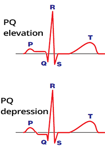
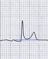
Ta Wave
|
|
Atrial Infarction
|
|
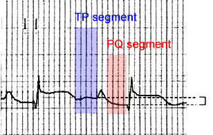
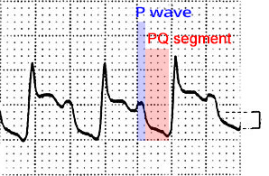
PQ (PTa) Segment
|
|
ECG and Atrial Infarction
|
|
|
PQ Depression (1mm)
|
PQ Depression (2mm)
|
|
Atrial Infarction
|
Pericarditis
|
Right Atrial Infarction and Inferior Wall STEMI
Acute Pericarditis
Right Atrial Infarction and STEMI of the Inferior Wall
Right Atrial Infarction and STEMI of the Inferior Wall
Atrial Infarction and Atrial Fibrillation
Sources