ECGbook.com

Making Medical Education Free for All

ECGbook.com

Making Medical Education Free for All

Home /

AV Dissociation

Atrioventricular (AV) dissociation

Sinus Rhythm

Sinus rhythm, SA node depolarization, electrical vector
ECG sinus rhythm, P wave, narrow QRS complex

Sinus Rhythm


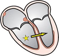
Ventricular Rhythm

Idioventricular rhythm, ectopic ventricular focus
ECG ventricular rhythm, broad QRS, regular rhythm

Accelerated Ventricular Rhythm
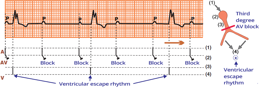

AV Dissociation

  • AV (Atrioventricular) Dissociation
  • Is independent activity of the atria and ventricles
  • There are 2 active pacemakers in the heart that generate impulses independently of each other
  • AV dissociation is not an arrhythmia
    • It is a symptom (sign) of some arrhythmia
      • AV dissociation always has a cause that led to it
    • Similarly, anemia is a symptom (sign) of some diagnosis
      • For example, bleeding from a stomach ulcer (diagnosis) causes blood loss (anemia)

Third-Degree AV Block (Complete AV Block)

Complete third (3rd) degree heart block

Laddergram (ECG) Complete 3rd degree AV block, atrioventricular (AV) dissociation

AV Dissociation in Third-Degree AV Block (Complete AV Block)

  • Laddergram illustrates the propagation of impulses through the conduction system
    • A - Atria, AV - AV junction, V - Ventricles
  • Third-Degree AV Block (Complete AV Block)
    • The atria and ventricles are electrically isolated from each other
      • There is a "cut-off" AV junction
    • Every impulse from the SA node (P wave) is blocked in the AV junction
  • AV Dissociation
    • There are 2 active pacemakers in the heart, which generate impulses independently

AV Dissociation - Classification


AV Dissociation - Causes


Third-degree atrioventricular block (AV block)
ECG 3rd degree complete AV block, First sinus rhythm (P waves), second ventricular rhythm (broad QRS)

Complete AV Dissociation


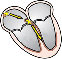
Incomplete atrioventricular dissociation, ventricular tachycardia, retrograde block
ECG incomplete av dissociation, ventricular tachycardia

Incomplete AV Dissociation

  • Initially there is a sinus rhythm
  • Ventricular extrasystole (3rd QRS) triggered ventricular tachycardia
    • AV junction is retrogradely depolarized from the ventricles
    • SA node generates impulses regularly (green arrows)
      • The ventricular focus generates impulses independently of the SA node
  • No impulse from the SA node reaches the ventricles
  • The patient does not have a "cut" AV junction


incomplete av dissociation, sa node depolarization, accelerated idioventricular rhythm
incomplete av dissociation, sinus rhythm and accelerated idioventricular rhythm

Incomplete AV Dissociation


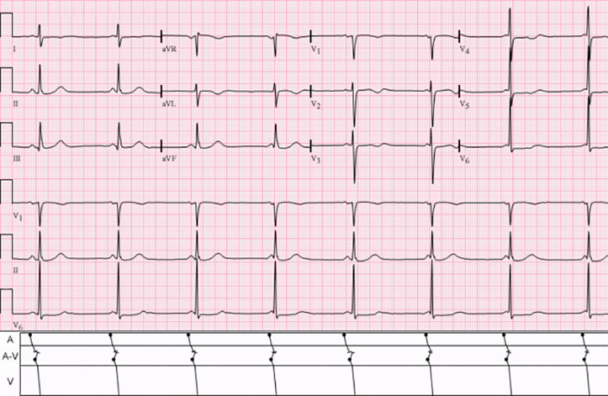
isorhythmic AV atrioventricular dissociation, SA node depolarization, accelerated idioventricular rhythm
ECG isorhythmic atrioventricular dissociation

Isorhythmic AV Dissociation
  • Laddergram (Rebríkový graf) illustrates impulse propagation through the conduction system
    • A - Atria, AV - AV junction, V - Ventricles
  • The heart has 2 pacemakers simultaneously
  • Both pacemakers have the same frequency
    • In each cycle, 2 impulses meet in the AV junction during the refractory period
    • The impulse from the SA node and the AV junction thus resets in the AV junction
  • No impulse from the SA node passes to the ventricles
    • In a longer ECG recording, a captured beat would definitely appear
      • In which the impulse from the SA node would pass to the ventricles
  • This is not a typical isorhythmic AV dissociation
    • Because the P waves (green rectangle) have a higher frequency than the QRS (orange stripe)
  • The patient does not have a "cut" AV junction


Isorhythmic atrioventricular (AV) dissociation, SA node depolarization, accelerated idioventricular rhythm
ECG isorhytmic AV dissociation, sinus rhythm, accelerated AV junctional rhythm

Isorhythmic AV Dissociation


Sources

  • ECG from Basics to Essentials Step by Step
  • litfl.com
  • ecgwaves.com
  • metealpaslan.com
  • medmastery.com
  • uptodate.com
  • ecgpedia.org
  • wikipedia.org
  • Strong Medicine
  • Understanding Pacemakers


šípka späť

AV Dissociation

Atrioventricular (AV) dissociation

Sinus Rhythm

Sinus rhythm, SA node depolarization, electrical vector

ECG sinus rhythm, P wave, narrow QRS complex

Sinus Rhythm


Ventricular Rhythm

Idioventricular rhythm, ectopic ventricular focus

ECG ventricular rhythm, broad QRS, regular rhythm

Accelerated Ventricular Rhythm


AV Dissociation

  • AV (Atrioventricular) Dissociation
  • Is independent activity of the atria and ventricles
  • There are 2 active pacemakers in the heart that generate impulses independently of each other
  • AV dissociation is not an arrhythmia
    • It is a symptom (sign) of some arrhythmia
      • AV dissociation always has a cause that led to it
    • Similarly, anemia is a symptom (sign) of some diagnosis
      • For example, bleeding from a stomach ulcer (diagnosis) causes blood loss (anemia)

Third-Degree AV Block (Complete AV Block)

  • The only electrical connection between the atria and the ventricles is the AV junction
  • In third-degree AV block, the AV junction is "cut off"
    • The atria and ventricles are electrically isolated from each other
    • Third-degree AV block is the most common cause of AV dissociation

  • The heart has 2 independent pacemakers in third-degree AV block:
    • 1st pacemaker (SA node, Sinus rhythm)
      • SA node generates impulses regularly (P waves)
      • Impulses from the SA node do not pass to the ventricles through the cut-off AV junction
    • 2nd pacemaker (Ventricular focus, Ventricular rhythm)
      • A focus activates spontaneously in the ventricles
        • This is a protective electrophysiological mechanism against asystole
      • The ventricular focus generates wide QRS complexes
        • QRS complexes occur independently of P waves (AV dissociation)

Complete third (3rd) degree heart block


Laddergram (ECG) Complete 3rd degree AV block, atrioventricular (AV) dissociation

AV Dissociation in Third-Degree AV Block (Complete AV Block)

  • Laddergram illustrates the propagation of impulses through the conduction system
    • A - Atria, AV - AV junction, V - Ventricles
  • Third-Degree AV Block (Complete AV Block)
    • The atria and ventricles are electrically isolated from each other
      • There is a "cut-off" AV junction
    • Every impulse from the SA node (P wave) is blocked in the AV junction
  • AV Dissociation
    • There are 2 active pacemakers in the heart, which generate impulses independently

AV Dissociation - Classification


AV Dissociation - Causes


ECG 3rd degree complete AV block, First sinus rhythm (P waves), second ventricular rhythm (broad QRS)

Complete AV Dissociation

  • P waves and QRS complexes are independent of each other
    • There are 2 pacemakers in the heart that generate impulses independently
  • The cause of AV dissociation is Third-Degree AV Block (Complete AV Block)
    • There is a "cut" AV junction
Third-degree atrioventricular block (AV block)


ECG incomplete av dissociation, ventricular tachycardia

Incomplete AV Dissociation

  • Initially there is a sinus rhythm
  • Ventricular extrasystole (3rd QRS) triggered ventricular tachycardia
    • AV junction is retrogradely depolarized from the ventricles
    • SA node generates impulses regularly (green arrows)
      • The ventricular focus generates impulses independently of the SA node
  • No impulse from the SA node reaches the ventricles
  • The patient does not have a "cut" AV junction
Incomplete atrioventricular dissociation, ventricular tachycardia, retrograde block


incomplete av dissociation, sinus rhythm and accelerated idioventricular rhythm

Incomplete AV Dissociation

  • The heart has 2 pacemakers simultaneously
  • Both pacemakers have the same frequency initially
    • In each cycle, 2 impulses meet in the AV junction during the refractory period
    • The impulses from the SA node and the ventricles thus reset in the AV junction
  • At the end of the ECG, the SA node overtakes the ventricular focus
    • The impulse from the SA node passes to the ventricles and depolarizes them (blue arrow)
    • After the P wave, a narrow QRS complex follows - blue arrow (sinus rhythm)
  • The patient does not have a "cut" AV junction
    • Does not have Third-Degree AV Block
    • At the end of the ECG, the impulse (P wave) passed to the ventricles (QRS) through the AV junction
  • The cause of AV dissociation is
incomplete av dissociation, sa node depolarization, accelerated idioventricular rhythm


ECG isorhythmic atrioventricular dissociation isorhythmic AV atrioventricular dissociation, SA node depolarization, accelerated idioventricular rhythm

Isorhythmic AV Dissociation

  • Laddergram (Rebríkový graf) illustrates impulse propagation through the conduction system
    • A - Atria, AV - AV junction, V - Ventricles
  • The heart has 2 pacemakers simultaneously
  • Both pacemakers have the same frequency
    • In each cycle, 2 impulses meet in the AV junction during the refractory period
    • The impulse from the SA node and the AV junction thus resets in the AV junction
  • No impulse from the SA node passes to the ventricles
    • In a longer ECG recording, a captured beat would definitely appear
      • In which the impulse from the SA node would pass to the ventricles
  • This is not a typical isorhythmic AV dissociation
    • Because the P waves (green rectangle) have a higher frequency than the QRS (orange stripe)
  • The patient does not have a "cut" AV junction


ECG isorhytmic AV dissociation, sinus rhythm, accelerated AV junctional rhythm

Isorhythmic AV Dissociation

  • Laddergram (Rebríkový graf) illustrates impulse propagation through the conduction system
    • A - Atria, AV - AV junction, V - Ventricles
  • The heart has 2 pacemakers simultaneously
  • Both pacemakers have the same frequency
    • In each cycle, 2 impulses meet in the AV junction during the refractory period
    • The impulse from the SA node and the AV junction thus resets in the AV junction
  • No impulse from the SA node passes to the ventricles
    • In a longer ECG recording, a captured beat would definitely appear
      • In which the impulse from the SA node would pass to the ventricles
  • The patient does not have a "cut" AV junction
Isorhythmic atrioventricular (AV) dissociation, SA node depolarization, accelerated idioventricular rhythm


Sources

  • ECG from Basics to Essentials Step by Step
  • litfl.com
  • ecgwaves.com
  • metealpaslan.com
  • medmastery.com
  • uptodate.com
  • ecgpedia.org
  • wikipedia.org
  • Strong Medicine
  • Understanding Pacemakers