ECGbook.com

Making Medical Education Free for All

ECGbook.com

Making Medical Education Free for All

Home /

Benign Early Repolarisation

Benign early repolarisation, J point elevation

Action Potential and Repolarization

ECG action potential phases, endocardium, epicardium depolarization, repolarization
  • Action Potential
    • Spreads in the myocardial wall from endocardium to epicardium
    • The action potential curve has a different shape in the endocardium and epicardium
      • Due to the different properties of ion channels
  • Ventricular Depolarization (Phase 0)
  • Ventricular Repolarization (Phase 1)
    • Begins on the ECG as the ST segment
    • Phase 1 is the beginning of repolarization
      • Often referred to as early repolarization

Benign Early Repolarization

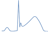
ECG action potential, J wave with notch, benign early repolarisation, repolarisation endocardium, epicardium

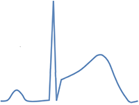
ECG and Benign Early Repolarization

ecg benign early repolarisation concave st elevation

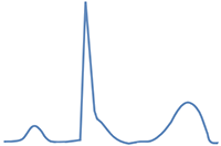
ecg convex st elevation
  • Elevation of the J-point in at least 2 adjacent leads
    • Most commonly in precordial leads (V2-6)
    • The J-point connects the QRS and ST segment
  • J wave in precordial leads
    • At the end of the QRS complex J wave
  • Concave ST elevations over the entire heart, but mainly precordial (V2-V6)
    • Most commonly up to 2mm in precordial leads, and up to 0.5mm in limb leads
    • Size depends on heart rate
  • Tall, peaked T waves over the entire heart, but mainly precordial (V2-V6)
  • ST/T ratio (V6) < 0.25
  • ST depression may be present (aVR)

  • Benign Early Repolarization (BER) is not found in patients over 50 years old

J-Point and Concave ST Elevations

ECG J point, ST elevation, ST segment

Variants of Benign Early Repolarization

  • BER can have the following 4 variants
    • Typically seen in 2 adjacent leads, most commonly in precordial leads (V2-6)
  • The first 2 variants are the most common
  • The second 2 variants without ST elevation are very rare

ECG classic benign early repolarization without a J wave ECG classic benign early repolarization with a J wave

Most Common BER Variants


ECG benign early repolarization slurred QRS downstroke without ST elevation ECG benign early repolarization J wave or the new J point elevation without ST elevation

Very Rare BER Variants


ECG J wave notch, fish hook pattern, benign early repolarisation

Fish Hook Pattern


Benign Early Repolarization vs. Pericarditis

ECG benign early repolarisation concave ST elevation
  • Benign Early Repolarization (BSR) and pericarditis both present with concave ST elevations
    • Differential diagnosis is made using additional ECG features


Pericarditis

  • ST elevations are in all leads (except aVR, V1)
  • PQ depressions are in all leads (except aVR, V1)
  • ST/T ratio (V6) > 0.25
  • No fish hook pattern (V4)
  • Normal T wave amplitude
  • ECG changes gradually resolve (approx. within 4 weeks)


Benign Early Repolarization

  • ST elevations are in the precordial leads (V2-V6)
  • Without PQ depressions
  • ST/T ratio (V6) < 0.25
  • Fish hook pattern (V4)
  • High amplitude T waves (T waves are symmetric)
  • ECG changes persist for years (do not resolve within 4 weeks)

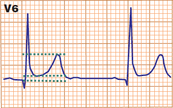
ST/T Ratio (V6)


ECG pericarditis ST/T ratio

Pericarditis


ECG benign early repolarisation ST/T ratio

Benign Early Repolarization


Inferolateral Early Repolarization

ECG inferior lateral benign early repolarisation, J wave notch
  • Benign Early Repolarization
    • is considered a benign diagnosis (a variant of normal)
    • If changes are observed only in the precordial leads (V2-V6)

  • Inferolateral Early Repolarization
    • Typically features a notch at the end of the QRS complex, found in at least two leads:
      • Inferior leads (II, III, aVF)
      • Lateral leads (I, aVL, V4-6)
    • According to some studies, it has a potentially increased risk:

Malignant Ventricular Arrhythmia


ECG benign early repolarisation, concave ST elevation, fish hook pattern, ST T ratio, J wave notch

Benign Early Repolarization

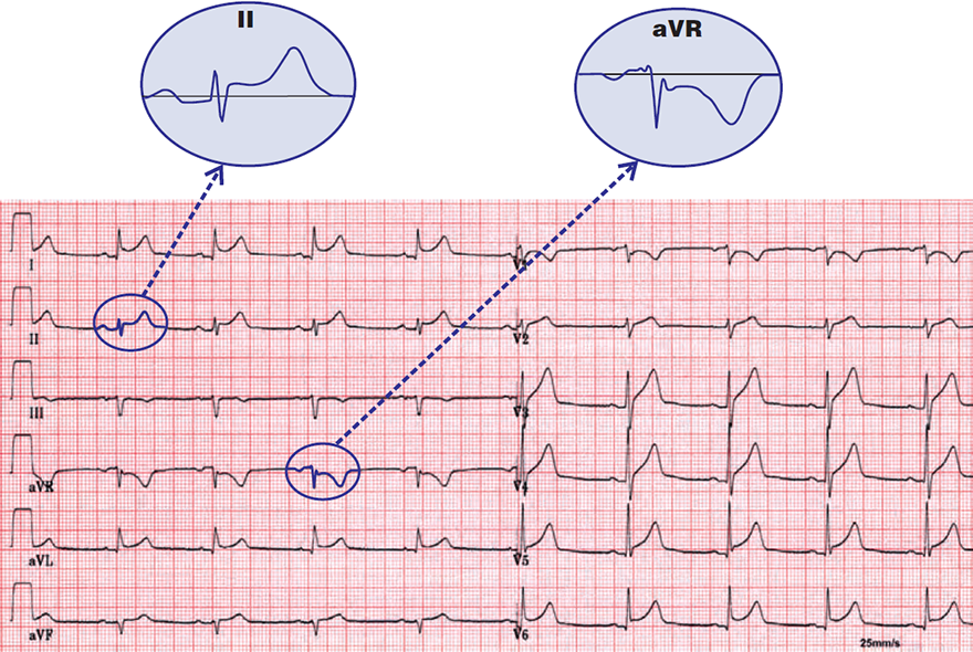
  • Concave ST elevations in precordial leads (V1-V6) and I
    • Pericarditis has ST elevations in all leads except (aVR, V1)
  • Fish-hook pattern (V3, V4, III)
    • Pericarditis does not have this fish-hook pattern
  • ST/T = 0.16
    • This indicates benign early repolarization
  • The patient has benign early repolarization


ECG pericarditis, concave ST elevation, spodick sign

Acute Pericarditis


ECG benign early repolarisation, J wave notch, fish hook pattern

Benign Early Repolarization

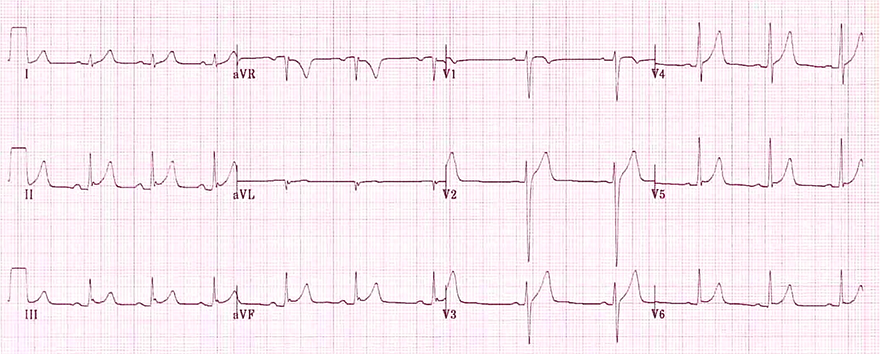
  • Concave ST elevations in almost all leads
    • Precordial (V2-V6) and inferolateral (I, II, III, aVF)
  • Fish-hook pattern (II, III, aVF)
  • High, peaked T waves (V2-V5, II)
  • ST/T = 0.16
    • This indicates benign early repolarization


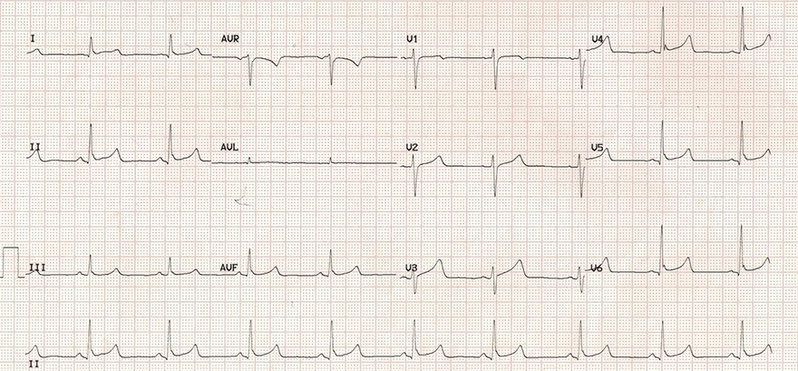
ECG benign early repolarisation, sinus bradycardia J wave

Benign Early Repolarization, Heart Rate 54/min


ECG benign early repolarisation, sinus rhythm

Benign Early Repolarization, Heart Rate 76/min

  • This is the same patient as on the previous ECG
  • Sinus Rhythm (76/min)
    • The ECG no longer shows sinus bradycardia
  • Signs of benign early repolarization disappear with increasing heart rate
    • Concave ST elevations have decreased
    • Fish-hook pattern has disappeared
  • This ECG does not show typical signs of BSR
    • These signs were present in the previous ECG with bradycardia


ECG benign early repolarisation, concave ST elevation, J wave, J point, peaked T waves, fish hook pattern

Benign Early Repolarization


ECG acute pericarditis, ST depression, PQ PR elevation aVR V1

Acute Pericarditis

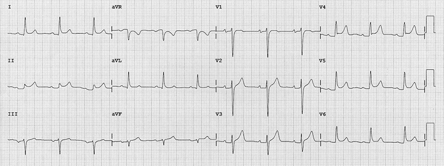
  • Concave ST elevations and PQ depressions in all leads except (aVR)
    • Benign early repolarization (BSR) does not have PQ depressions
  • No fish-hook pattern in precordial leads
  • ST/T = 0.33
    • Indicating pericarditis
  • Patient has pericarditis (stage 1)


ECG acute pericarditis 1 stage, concave ST elevation, PQ PR depression, sinus tachycardia

Acute Pericarditis


Sources

  • ECG from Basics to Essentials Step by Step
  • litfl.com
  • ecgwaves.com
  • metealpaslan.com
  • medmastery.com
  • uptodate.com
  • ecgpedia.org
  • wikipedia.org
  • Strong Medicine
  • Understanding Pacemakers


šípka späť

Benign Early Repolarisation

Benign early repolarisation, J point elevation

Action Potential and Repolarization

ECG action potential phases, endocardium, epicardium depolarization, repolarization
  • Action Potential
    • Spreads in the myocardial wall from endocardium to epicardium
    • The action potential curve has a different shape in the endocardium and epicardium
      • Due to the different properties of ion channels
  • Ventricular Depolarization (Phase 0)
  • Ventricular Repolarization (Phase 1)
    • Begins on the ECG as the ST segment
    • Phase 1 is the beginning of repolarization
      • Often referred to as early repolarization

Benign Early Repolarization

ECG action potential, J wave with notch, benign early repolarisation, repolarisation endocardium, epicardium
  • It is a benign ion channel disturbance in the epicardium
    • occurring during early repolarization (Phase 1)
  • On the ECG it appears as
    • Concave ST elevation
    • Higher ST segment elevation
    • Notch (J wave)

  • Prevalence is approximately 5% of the population
  • Considered a variant of normal
  • Typically found in young people under 50 years old, especially:

ECG and Benign Early Repolarization

  • Elevation of the J-point in at least 2 adjacent leads
    • Most commonly in precordial leads (V2-6)
    • The J-point connects the QRS and ST segment
  • J wave in precordial leads
    • At the end of the QRS complex J wave
  • Concave ST elevations over the entire heart, but mainly precordial (V2-V6)
    • Most commonly up to 2mm in precordial leads, and up to 0.5mm in limb leads
    • Size depends on heart rate
  • Tall, peaked T waves over the entire heart, but mainly precordial (V2-V6)
  • ST/T ratio (V6) < 0.25
  • ST depression may be present (aVR)

  • Benign Early Repolarization (BER) is not found in patients over 50 years old

ecg benign early repolarisation concave st elevation

ecg convex st elevation

J-Point and Concave ST Elevations

  • J-Point and Concave (Smiling) ST Elevations
    • Are key for diagnosing Benign Early Repolarization (BER)
  • The J-Point connects the QRS and ST segment
    • In cases of J wave and a creeping QRS complex, the position of the J-point is debatable

ECG J point, ST elevation, ST segment

Variants of Benign Early Repolarization

  • BER can have the following 4 variants
    • Typically seen in 2 adjacent leads, most commonly in precordial leads (V2-6)
  • The first 2 variants are the most common
  • The second 2 variants without ST elevation are very rare


ECG classic benign early repolarization without a J wave ECG classic benign early repolarization with a J wave ECG benign early repolarization slurred QRS downstroke without ST elevation ECG benign early repolarization J wave or the new J point elevation without ST elevation

Most Common BER Variants

Very Rare BER Variants
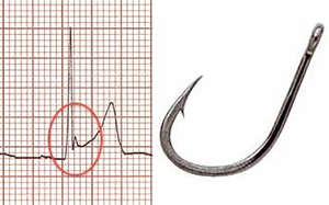


ECG J wave notch, fish hook pattern, benign early repolarisation

Fish Hook Pattern

  • J wave and concave ST elevation resemble a fish hook
  • Most clearly visible in V4
  • Almost always present in Benign Early Repolarization
  • Pericarditis does not have this pattern

Benign Early Repolarization vs. Pericarditis

  • Benign Early Repolarization (BSR) and pericarditis both present with concave ST elevations
    • Differential diagnosis is made using additional ECG features

ECG benign early repolarisation concave ST elevation

Pericarditis

  • ST elevations are in all leads (except aVR, V1)
  • PQ depressions are in all leads (except aVR, V1)
  • ST/T ratio (V6) > 0.25
  • No fish hook pattern (V4)
  • Normal T wave amplitude
  • ECG changes gradually resolve (approx. within 4 weeks)

Benign Early Repolarization

  • ST elevations are in the precordial leads (V2-V6)
  • Without PQ depressions
  • ST/T ratio (V6) < 0.25
  • Fish hook pattern (V4)
  • High amplitude T waves (T waves are symmetric)
  • ECG changes persist for years (do not resolve within 4 weeks)

ST/T Ratio (V6)


ECG pericarditis ST/T ratio ECG benign early repolarisation ST/T ratio

Pericarditis

  • ST elevation = 1.5mm
  • T wave = 3.5mm
  • ST/T = 0.43
    • ST/T > 0.25 (Pericarditis)

Benign Early Repolarization

  • ST elevation = 1mm
  • T wave = 6mm
  • ST/T = 0.16
    • ST/T < 0.25 (Benign Early Repolarization)

Inferolateral Early Repolarization

  • Benign Early Repolarization
    • is considered a benign diagnosis (a variant of normal)
    • If changes are observed only in the precordial leads (V2-V6)

  • Inferolateral Early Repolarization
    • Typically features a notch at the end of the QRS complex, found in at least two leads:
      • Inferior leads (II, III, aVF)
      • Lateral leads (I, aVL, V4-6)
    • According to some studies, it has a potentially increased risk:


ECG inferior lateral benign early repolarisation, J wave notch

Malignant Ventricular Arrhythmia


ECG benign early repolarisation, concave ST elevation, fish hook pattern, ST T ratio, J wave notch

Benign Early Repolarization

  • Concave ST elevations in precordial leads (V1-V6) and I
    • Pericarditis has ST elevations in all leads except (aVR, V1)
  • Fish-hook pattern (V3, V4, III)
    • Pericarditis does not have this fish-hook pattern
  • ST/T = 0.16
    • This indicates benign early repolarization
  • The patient has benign early repolarization


ECG pericarditis, concave ST elevation, spodick sign

Acute Pericarditis


ECG benign early repolarisation, J wave notch, fish hook pattern

Benign Early Repolarization

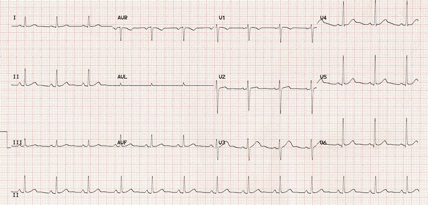
  • Concave ST elevations in almost all leads
    • Precordial (V2-V6) and inferolateral (I, II, III, aVF)
  • Fish-hook pattern (II, III, aVF)
  • High, peaked T waves (V2-V5, II)
  • ST/T = 0.16
    • This indicates benign early repolarization


ECG benign early repolarisation, sinus bradycardia J wave

Benign Early Repolarization, Heart Rate 54/min


ECG benign early repolarisation, sinus rhythm

Benign Early Repolarization, Heart Rate 76/min
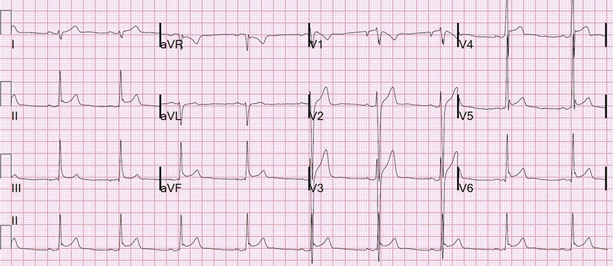
  • This is the same patient as on the previous ECG
  • Sinus Rhythm (76/min)
    • The ECG no longer shows sinus bradycardia
  • Signs of benign early repolarization disappear with increasing heart rate
    • Concave ST elevations have decreased
    • Fish-hook pattern has disappeared
  • This ECG does not show typical signs of BSR
    • These signs were present in the previous ECG with bradycardia


ECG benign early repolarisation, concave ST elevation, J wave, J point, peaked T waves, fish hook pattern

Benign Early Repolarization


ECG acute pericarditis, ST depression, PQ PR elevation aVR V1

Acute Pericarditis

  • Concave ST elevations and PQ depressions in all leads except (aVR)
    • Benign early repolarization (BSR) does not have PQ depressions
  • No fish-hook pattern in precordial leads
  • ST/T = 0.33
    • Indicating pericarditis
  • Patient has pericarditis (stage 1)


ECG acute pericarditis 1 stage, concave ST elevation, PQ PR depression, sinus tachycardia

Acute Pericarditis


Sources

  • ECG from Basics to Essentials Step by Step
  • litfl.com
  • ecgwaves.com
  • metealpaslan.com
  • medmastery.com
  • uptodate.com
  • ecgpedia.org
  • wikipedia.org
  • Strong Medicine
  • Understanding Pacemakers