Home /
Concealed Conduction
Concealed conduction, Concealed extrasystoles, Pseudo AV block, Reciprocal Echo beat
Conduction System and Concealed Conduction

- Impulse (action potential) propagates through
- Electrical vector in different parts of the heart
- Concealed conduction
- Is impulse conduction through a certain part of the conduction system
- that is not visible on the ECG curve (is concealed)
- We diagnose it based on the subsequent ECG curve
- Concealed conduction most commonly manifests in the AV junction
Mechanism of Concealed Conduction
- Concealed conduction most commonly manifests in the AV junction
- If an impulse penetrates the AV junction at a specific time (most often retrogradely from a ventricular extrasystole)
- Then the AV junction conducts the next impulse more slowly
- The principle is a timed electrophysiological modification of the refractory period
Ventricular Extrasystole and Concealed Conduction

Concealed Conduction and Ventricular Extrasystole
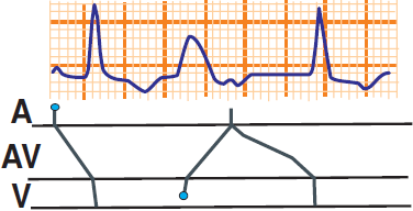
- A laddergram illustrates impulse propagation through the conduction system
- A - Atria, AV - AV junction, V - Ventricles
- (1) An interpolated ventricular extrasystole (VES) - occurs between 2 sinus beats
- The impulse from the VES retrogradely penetrates the AV junction and alters its electrophysiological properties
- The following sinus impulse reaches the AV junction during the partial refractory period
- The impulse conducts more slowly through the AV junction
- Which is seen on the ECG as a prolonged PQ interval
- Concealed conduction is the retrograde conduction of the VES into the AV junction, which is not visible on the ECG
- We only see the subsequent prolonged PQ interval
- (2) Ventricular Extrasystole
- Retrogradely penetrates the AV junction
- The following sinus impulse reaches the AV junction during the refractory period and gets blocked

Concealed Conduction and Ventricular Extrasystole
- Sinus rhythm with a PQ interval of 0.18s
- Two interpolated ventricular extrasystoles (VES)
- Each followed by a prolonged PQ interval of 0.32s
- The retrograde impulse from the VES penetrates the AV junction
- And slows the subsequent conduction of the sinus impulse
- Which is seen as a prolonged PQ interval of 0.32s (after VES)

Ventricular Extrasystole (VES)
- The retrograde impulse from the VES penetrated the AV junction
- The P wave after the VES is blocked
- In the AV junction, the sinus and retrograde impulses from the VES met in the refractory phase, blocking each other
- After the VES, a complete compensatory pause follows
- 2x the RR interval without VES = RR interval with VES
- Concealed conduction is not present
Atrial Fibrillation and Concealed Conduction
- In atrial fibrillation, impulses in the atria occur irregularly with a frequency of 350-600/min.
- These impulses are filtered in the AV junction, resulting in an irregularly irregular ventricular rate
- The AV junction acts as a filter, allowing, for example, every fourth impulse to pass to the ventricles
- After an impulse passes through the AV junction, a refractory period occurs
- This temporarily blocks subsequent impulses
- If an impulse reaches the AV junction at a specific time
- It results in concealed conduction (partial refractory period) and the next impulse is conducted more slowly
- Concealed conduction during atrial fibrillation is not visible on the ECG because the PQ interval cannot be differentiated

Concealed Conduction and Atrial Fibrillation
- Supraventricular impulses in atrial fibrillation
- Some impulses create concealed conduction (the next impulse is conducted more slowly)
- Which can be seen on the laddergram in the AV part as a different angle of AV conduction
- It is not visible on the ECG because, in atrial fibrillation, it is impossible to differentiate the PQ interval
Pseudo AV Block and Concealed Conduction
- Concealed conduction can sometimes mimic an AV block on the ECG
- However, this is a pseudo AV block with a different electrophysiological mechanism

Concealed Conduction and Pseudo AV Block II Degree (Mobitz II)
- The ECG shows AV block II degree (Mobitz II)
- Every 2nd P wave is blocked
- This is concealed conduction that mimics an AV block on the ECG
- In a long-term ECG recording, the ECG pattern of AV block II degree will not continue
- Because the junctional extrasystole would have to occur exactly at the time
- when the AV junction is in the refractory phase due to the impulse from the SA node
- This is impossible in a long-term ECG recording

Concealed Conduction and Pseudo AV Block of the First Degree
- The ECG shows an AV block of the first degree
- It is characterized by a prolonged PQ interval > 0.2s
- This is concealed conduction that mimics an AV block on the ECG
- Junctional extrasystole and a sinus impulse meet in the AV junction during sinus rhythm
- The junctional extrasystole partially penetrates the AV junction, causing the SA impulse to be conducted more slowly through the AV junction
- This results in a partial refractory period in the AV junction, which alters the conduction speed of the following impulses
- The PQ interval is prolonged
- In a long-term ECG recording, the ECG pattern of AV block of the first degree will not persist
- Due to the unlikely repeated timing of the junctional extrasystole
Reciprocal Echo Beat and Concealed Conduction
- The AV junction has 2 pathways (slow and fast)
- Reciprocal Echo Beat
- Occurs with specific timing of an extrasystole, when the impulse "bounces off" the AV junction
- The impulse revolves around the AV junction (through the 2 pathways) so that it does not encounter its own refractory period
- One impulse thus activates both the atria and ventricles from the AV junction

Concealed Conduction and Reciprocal Echo Beats
- This is concealed conduction, where one impulse creates multiple ECG waveforms (P waves, QRS complexes)
- Atrial Echo
- The impulse from an atrial extrasystole
- Revolves around the AV junction
- One impulse activates the atria twice and the ventricles once
- Junctional Echo
- The impulse from a junctional extrasystole
- Revolves around the AV junction
- One impulse activates the atria once and the ventricles twice
- Ventricular Echo
- The impulse from a ventricular extrasystole
- Revolves around the AV junction
- One impulse activates the atria once and the ventricles twice

Concealed Conduction and Pseudo AV Block of the First Degree
- On the ECG, there is a ventricular echo beat
- However, with a long ECG recording, the ECG pattern of first-degree AV block will not persist
- Due to the unlikely repeated timing of the ventricular extrasystole

Atrial Echo and Second-Degree AV Block (Wenckebach)
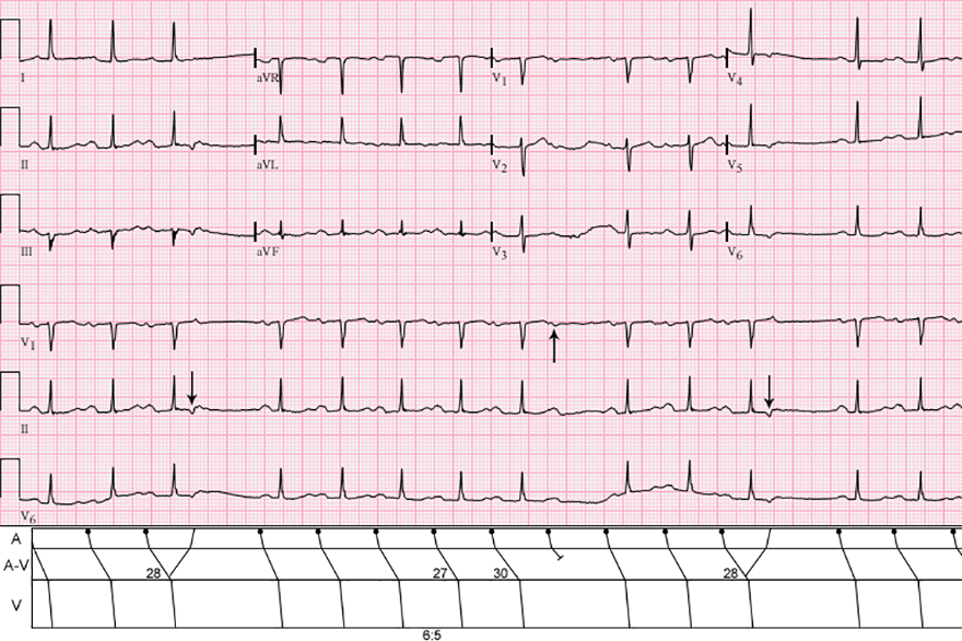
- On the ECG, there is second-degree AV block (Mobitz I - Wenckebach) with variable conduction to the ventricles
- PQ interval progressively lengthens until the P wave is blocked
- In the middle of the ECG recording, the ventricular conduction ratio is 6:5 (6th P wave is blocked)
- Notice on the laddergram, the delay of impulse in the AV node is 27ms, 28ms, 30ms
- For the impulse to turn around in the AV junction, it must not encounter the refractory period (must be precisely timed)
- Atrial Echo occurs only when
- The conduction in the AV node is extended to 28ms
- Then the impulse turns around in the AV junction and does not encounter the refractory period
Sources
- ECG from Basics to Essentials Step by Step
- litfl.com
- ecgwaves.com
- metealpaslan.com
- medmastery.com
- uptodate.com
- ecgpedia.org
- wikipedia.org
- Strong Medicine
- Understanding Pacemakers

Home /
Concealed Conduction
Concealed conduction, Concealed extrasystoles, Pseudo AV block, Reciprocal Echo beat
Conduction System and Concealed Conduction
- Impulse (action potential) propagates through
- Electrical vector in different parts of the heart
- Concealed conduction
- Is impulse conduction through a certain part of the conduction system
- that is not visible on the ECG curve (is concealed)
- We diagnose it based on the subsequent ECG curve
- Concealed conduction most commonly manifests in the AV junction
|

|
Mechanism of Concealed Conduction
- Concealed conduction most commonly manifests in the AV junction
- If an impulse penetrates the AV junction at a specific time (most often retrogradely from a ventricular extrasystole)
- Then the AV junction conducts the next impulse more slowly
- The principle is a timed electrophysiological modification of the refractory period
Ventricular Extrasystole and Concealed Conduction

Concealed Conduction and Ventricular Extrasystole
- A laddergram illustrates impulse propagation through the conduction system
- A - Atria, AV - AV junction, V - Ventricles
- (1) An interpolated ventricular extrasystole (VES) - occurs between 2 sinus beats
- The impulse from the VES retrogradely penetrates the AV junction and alters its electrophysiological properties
- The following sinus impulse reaches the AV junction during the partial refractory period
- The impulse conducts more slowly through the AV junction
- Which is seen on the ECG as a prolonged PQ interval
- Concealed conduction is the retrograde conduction of the VES into the AV junction, which is not visible on the ECG
- We only see the subsequent prolonged PQ interval
- (2) Ventricular Extrasystole
- Retrogradely penetrates the AV junction
- The following sinus impulse reaches the AV junction during the refractory period and gets blocked

Concealed Conduction and Ventricular Extrasystole
- Sinus rhythm with a PQ interval of 0.18s
- Two interpolated ventricular extrasystoles (VES)
- Each followed by a prolonged PQ interval of 0.32s
- The retrograde impulse from the VES penetrates the AV junction
- And slows the subsequent conduction of the sinus impulse
- Which is seen as a prolonged PQ interval of 0.32s (after VES)

Ventricular Extrasystole (VES)
- The retrograde impulse from the VES penetrated the AV junction
- The P wave after the VES is blocked
- In the AV junction, the sinus and retrograde impulses from the VES met in the refractory phase, blocking each other
- After the VES, a complete compensatory pause follows
- 2x the RR interval without VES = RR interval with VES
- Concealed conduction is not present
Atrial Fibrillation and Concealed Conduction
- In atrial fibrillation, impulses in the atria occur irregularly with a frequency of 350-600/min.
- These impulses are filtered in the AV junction, resulting in an irregularly irregular ventricular rate
- The AV junction acts as a filter, allowing, for example, every fourth impulse to pass to the ventricles
- After an impulse passes through the AV junction, a refractory period occurs
- This temporarily blocks subsequent impulses
- If an impulse reaches the AV junction at a specific time
- It results in concealed conduction (partial refractory period) and the next impulse is conducted more slowly
- Concealed conduction during atrial fibrillation is not visible on the ECG because the PQ interval cannot be differentiated

Concealed Conduction and Atrial Fibrillation
- Supraventricular impulses in atrial fibrillation
- Some impulses create concealed conduction (the next impulse is conducted more slowly)
- Which can be seen on the laddergram in the AV part as a different angle of AV conduction
- It is not visible on the ECG because, in atrial fibrillation, it is impossible to differentiate the PQ interval
Pseudo AV Block and Concealed Conduction
- Concealed conduction can sometimes mimic an AV block on the ECG
- However, this is a pseudo AV block with a different electrophysiological mechanism

Concealed Conduction and Pseudo AV Block II Degree (Mobitz II)
- The ECG shows AV block II degree (Mobitz II)
- Every 2nd P wave is blocked
- This is concealed conduction that mimics an AV block on the ECG
- In a long-term ECG recording, the ECG pattern of AV block II degree will not continue
- Because the junctional extrasystole would have to occur exactly at the time
- when the AV junction is in the refractory phase due to the impulse from the SA node
- This is impossible in a long-term ECG recording

Concealed Conduction and Pseudo AV Block of the First Degree
- The ECG shows an AV block of the first degree
- It is characterized by a prolonged PQ interval > 0.2s
- This is concealed conduction that mimics an AV block on the ECG
- Junctional extrasystole and a sinus impulse meet in the AV junction during sinus rhythm
- The junctional extrasystole partially penetrates the AV junction, causing the SA impulse to be conducted more slowly through the AV junction
- This results in a partial refractory period in the AV junction, which alters the conduction speed of the following impulses
- The PQ interval is prolonged
- In a long-term ECG recording, the ECG pattern of AV block of the first degree will not persist
- Due to the unlikely repeated timing of the junctional extrasystole
Reciprocal Echo Beat and Concealed Conduction
- The AV junction has 2 pathways (slow and fast)
- Reciprocal Echo Beat
- Occurs with specific timing of an extrasystole, when the impulse "bounces off" the AV junction
- The impulse revolves around the AV junction (through the 2 pathways) so that it does not encounter its own refractory period
- One impulse thus activates both the atria and ventricles from the AV junction

Concealed Conduction and Reciprocal Echo Beats
- This is concealed conduction, where one impulse creates multiple ECG waveforms (P waves, QRS complexes)
- Atrial Echo
- The impulse from an atrial extrasystole
- Revolves around the AV junction
- One impulse activates the atria twice and the ventricles once
- Junctional Echo
- The impulse from a junctional extrasystole
- Revolves around the AV junction
- One impulse activates the atria once and the ventricles twice
- Ventricular Echo
- The impulse from a ventricular extrasystole
- Revolves around the AV junction
- One impulse activates the atria once and the ventricles twice

Concealed Conduction and Pseudo AV Block of the First Degree
- On the ECG, there is a ventricular echo beat
- However, with a long ECG recording, the ECG pattern of first-degree AV block will not persist
- Due to the unlikely repeated timing of the ventricular extrasystole

Atrial Echo and Second-Degree AV Block (Wenckebach)
- On the ECG, there is second-degree AV block (Mobitz I - Wenckebach) with variable conduction to the ventricles
- PQ interval progressively lengthens until the P wave is blocked
- In the middle of the ECG recording, the ventricular conduction ratio is 6:5 (6th P wave is blocked)
- Notice on the laddergram, the delay of impulse in the AV node is 27ms, 28ms, 30ms
- For the impulse to turn around in the AV junction, it must not encounter the refractory period (must be precisely timed)
- Atrial Echo occurs only when
- The conduction in the AV node is extended to 28ms
- Then the impulse turns around in the AV junction and does not encounter the refractory period
Sources
- ECG from Basics to Essentials Step by Step
- litfl.com
- ecgwaves.com
- metealpaslan.com
- medmastery.com
- uptodate.com
- ecgpedia.org
- wikipedia.org
- Strong Medicine
- Understanding Pacemakers


