|
|
ECGbook.com Making Medical Education Free for All |
|
|
ECGbook.com Making Medical Education Free for All |
|
|
ECGbook.com Making Medical Education Free for All |
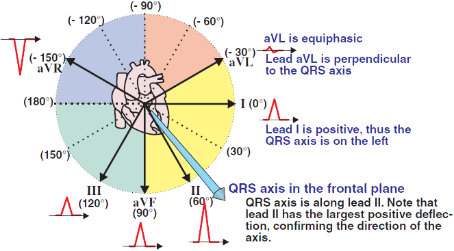
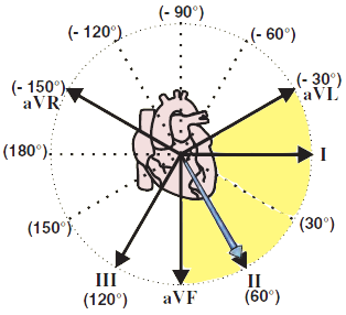
Main Cardiac Vector and the 4 Quadrants
Magnitude of the R Wave and the Main Cardiac Vector
Biphasic QRS Complex
Normal (Intermediate) Cardiac Axis
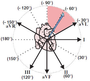
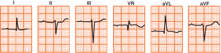
Cardiac Axis in the Upper Left Quadrant
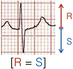
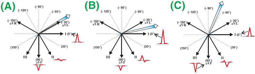
Equiphasic
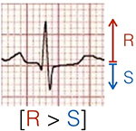
Predominantly Positive
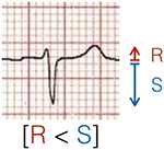
Predominantly Negative
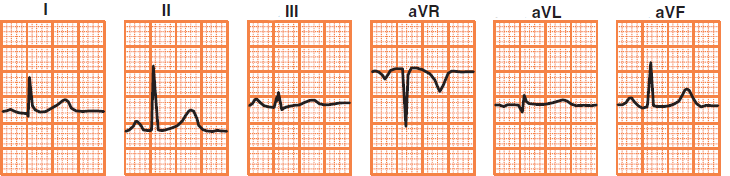
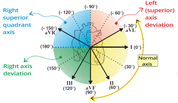
Normal Cardiac Axis
Left Axis Deviation
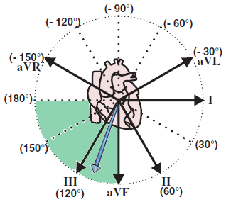
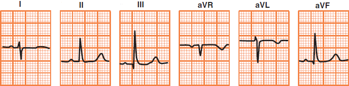
Right Axis Deviation
Extreme Axis Deviation
Sources
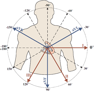
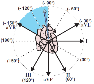
Limb ECG Leads
|
|
|
|
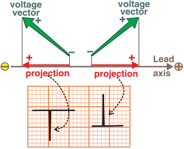
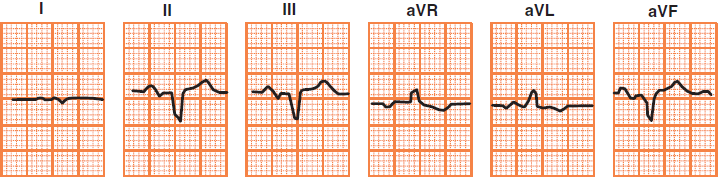
Positive and Negative ECG Deflections
|
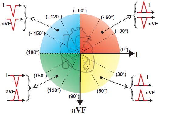
Main Cardiac Vector and the 4 Quadrants
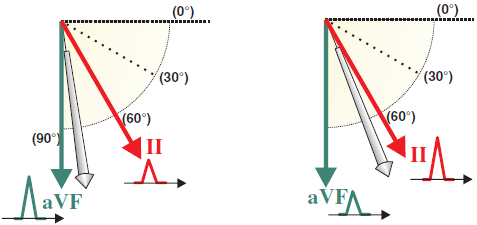
Magnitude of the R Wave and the Main Cardiac Vector
Biphasic QRS Complex
Normal (Intermediate) Cardiac Axis
Cardiac Axis in the Upper Left Quadrant
|
Equiphasic
|
Predominantly Positive
|
Predominantly Negative
|
|
|
|
|
Normal Cardiac Axis
|
|
|
|
|
|
Left Axis Deviation
|
|
|
|
|
|
Right Axis Deviation
|
|
|
|
|
|
Extreme Axis Deviation
|
|
Sources