ECGbook.com

Making Medical Education Free for All

ECGbook.com

Making Medical Education Free for All

Home /

Dextrocardia

Dextrocardia

Dextrocardia and X-ray

x-ray-dextrocardia-image
  • Levocardia
    • It is the physiological position of the heart, with the apex of the heart pointing to the left
  • Dextroposition
    • The entire heart is shifted to the right in the chest
    • The apex of the heart still points to the left
  • Dextrocardia
    • It is a congenital developmental defect of the heart
    • The position of the heart base is shifted only minimally
    • The apex of the heart points to the right
  • A chest X-ray is sufficient for diagnosing dextrocardia
    • No ECG is needed

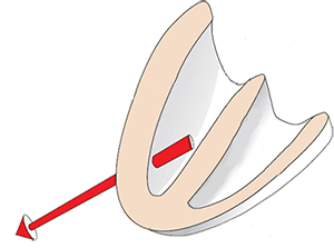
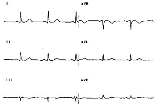
Main Cardiac Vector

Dextrocardia and direction of heart electrical vector

ECG and Decreased R Wave Progression

  • In levocardia
    • The main vector points to V5-V6
    • R wave increases from V1 to V5
  • In dextrocardia
    • The main vector points away from V5-V6
    • R wave does not increase from V1 to V5


  • Decreased R wave progression is:
    • R wave in V3 < 4mm
      • The R wave does not increase from V1 to V5
    • or amputated R waves
      • R wave (V1-V3) is absent
ECG R wave progression from V1 to V5

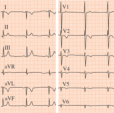
ECG poor R wave progression, R wave in V3 less than 4mm

ECG and Dextrocardia

ECG dextrocardia criteria: inverted I lead, poor r wave progression,

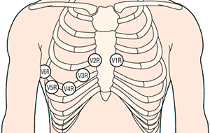
Mirror Chest Electrodes

Dextrocardia diagnosis with ecg right sided leads (V1R-V6R)
  • In case of suspicion of dextrocardia (for extreme axis deviation to the right)
    • Mirror chest electrodes (V1R-V6R) are placed

  • If normal R wave progression appears in the mirror leads
    • It indicates dextrocardia

Reversed Arm Electrodes vs. Dextrocardia


ECG differential diagnosis dextrocardia vs. reversal of the left and right arm electrodes

Differential Diagnosis: Dextrocardia vs. Reversed Arm Electrodes
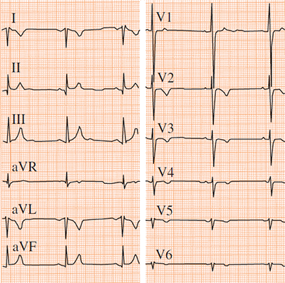


ECG dextrocardia with poor R wave progression

Dextrocardia


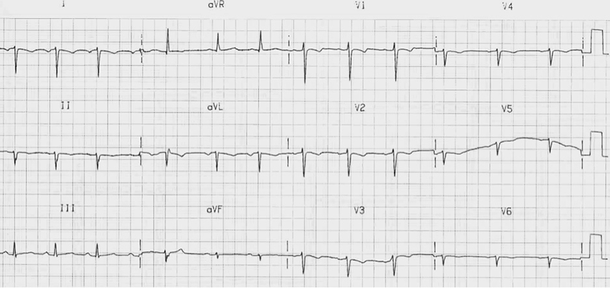
ECG reversal of the left and right arm electrodes, with normal R wave progression

Reversed Arm Electrodes

  • Extreme Right Axis Deviation
    • Dominant R in aVR
    • Dominant S in Lead I
  • R Wave Progression
    • R wave from V1 to V6 increases


ECG dextrocardia reversal lead I, and poor r wave progression

Dextrocardia


ECG with reveral limb leads right arm and left arm with normal r wave progression and reversal I lead

Reversed Arm Electrodes

  • Extreme Right Axis Deviation
    • Dominant R in aVR
    • Dominant S in Lead I
  • R Wave Progression
    • R wave from V1 to V6 increases
  • The patient has reversed limb electrodes (left arm/right arm)


ECG with correct lieb leads (right arm, left arm) position and with normal r wave progression, and with positive lead I

Correctly Placed Arm Electrodes


ECG dextrocardia with old inferior STEMI infarction. Poor r wave progression

Dextrocardia and Old Inferior Myocardial Infarction


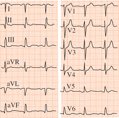
ECG Right sided precordial leads, old inferior stemi, dextrocardia with poor r wave progression

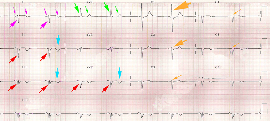
Right-Sided Chest Leads (V1R-V6R)

12-lead ECG with Right sided precordial leads


Sources

  • ECG from Basics to Essentials Step by Step
  • litfl.com
  • ecgwaves.com
  • metealpaslan.com
  • medmastery.com
  • uptodate.com
  • ecgpedia.org
  • wikipedia.org
  • Strong Medicine
  • Understanding Pacemakers


šípka späť

Dextrocardia

Dextrocardia

Dextrocardia and X-ray

  • Levocardia
    • It is the physiological position of the heart, with the apex of the heart pointing to the left
  • Dextroposition
    • The entire heart is shifted to the right in the chest
    • The apex of the heart still points to the left
  • Dextrocardia
    • It is a congenital developmental defect of the heart
    • The position of the heart base is shifted only minimally
    • The apex of the heart points to the right
  • A chest X-ray is sufficient for diagnosing dextrocardia
    • No ECG is needed

x-ray-dextrocardia-image

Main Cardiac Vector


Dextrocardia and direction of heart electrical vector

ECG and Decreased R Wave Progression

  • In levocardia
    • The main vector points to V5-V6
    • R wave increases from V1 to V5
  • In dextrocardia
    • The main vector points away from V5-V6
    • R wave does not increase from V1 to V5


  • Decreased R wave progression is:
    • R wave in V3 < 4mm
      • The R wave does not increase from V1 to V5
    • or amputated R waves
      • R wave (V1-V3) is absent
ECG R wave progression from V1 to V5

ECG poor R wave progression, R wave in V3 less than 4mm

ECG and Dextrocardia


ECG dextrocardia criteria: inverted I lead, poor r wave progression,

Mirror Chest Electrodes

  • In case of suspicion of dextrocardia (for extreme axis deviation to the right)
    • Mirror chest electrodes (V1R-V6R) are placed

  • If normal R wave progression appears in the mirror leads
    • It indicates dextrocardia

Dextrocardia diagnosis with ecg right sided leads (V1R-V6R)

Reversed Arm Electrodes vs. Dextrocardia


ECG differential diagnosis dextrocardia vs. reversal of the left and right arm electrodes

Differential Diagnosis: Dextrocardia vs. Reversed Arm Electrodes


ECG dextrocardia with poor R wave progression ECG reversal of the left and right arm electrodes, with normal R wave progression

Dextrocardia

Reversed Arm Electrodes
  • Extreme Right Axis Deviation
    • Dominant R in aVR
    • Dominant S in Lead I
  • R Wave Progression
    • R wave from V1 to V6 increases


ECG dextrocardia reversal lead I, and poor r wave progression

Dextrocardia


ECG with reveral limb leads right arm and left arm with normal r wave progression and reversal I lead

Reversed Arm Electrodes

  • Extreme Right Axis Deviation
    • Dominant R in aVR
    • Dominant S in Lead I
  • R Wave Progression
    • R wave from V1 to V6 increases
  • The patient has reversed limb electrodes (left arm/right arm)


ECG with correct lieb leads (right arm, left arm) position and with normal r wave progression, and with positive lead I

Correctly Placed Arm Electrodes


ECG dextrocardia with old inferior STEMI infarction. Poor r wave progression

Dextrocardia and Old Inferior Myocardial Infarction
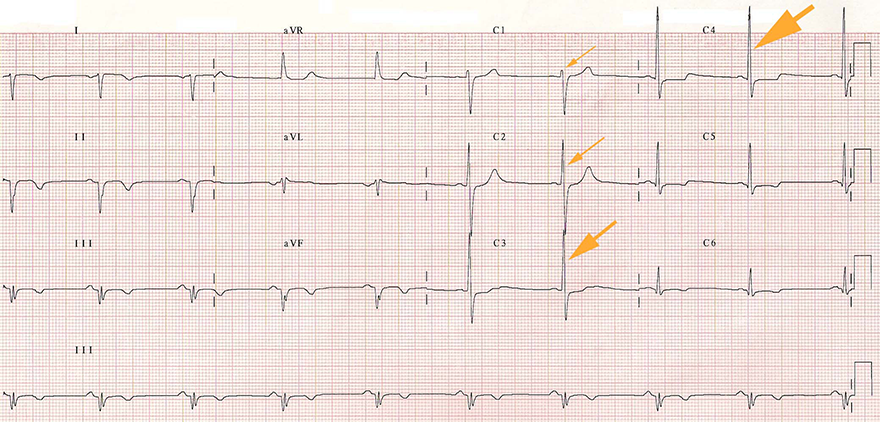


ECG Right sided precordial leads, old inferior stemi, dextrocardia with poor r wave progression

Right-Sided Chest Leads (V1R-V6R)

  • This is an ECG from a previous patient
  • The chest leads are mirror-image placed on the right
    • They are labeled (V1R-V6R)
    • The ECG shows R wave progression from V1 to V6
    • The limb leads remain unchanged
  • The ECG shows R wave progression (brown-yellow arrows)
    • Because the vector points to the right towards leads VR5-VR6
12-lead ECG with Right sided precordial leads


Sources

  • ECG from Basics to Essentials Step by Step
  • litfl.com
  • ecgwaves.com
  • metealpaslan.com
  • medmastery.com
  • uptodate.com
  • ecgpedia.org
  • wikipedia.org
  • Strong Medicine
  • Understanding Pacemakers