|
|
ECGbook.com Making Medical Education Free for All |
Upload ECG for Interpretation |
|
|
ECGbook.com Making Medical Education Free for All |
Upload ECG for Interpretation |
|
|
ECGbook.com Making Medical Education Free for All |
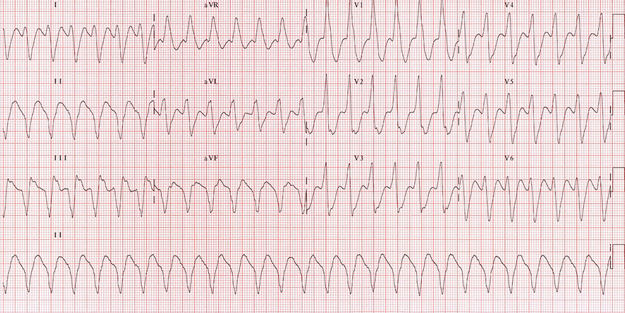
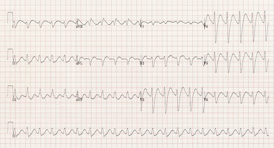
Ventricular Tachycardia
Tricyclic Antidepressants (Intoxication)

Dextrocardia
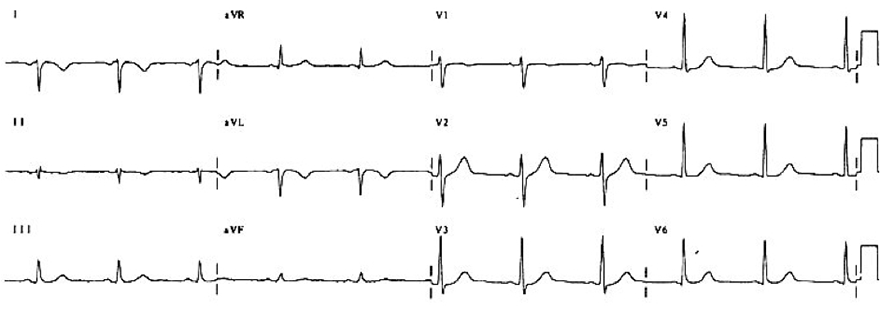
Swapped ECG Leads (Left and Right Arm)
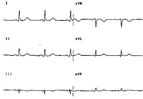
Correctly Placed Limb ECG Leads
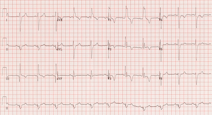
Acute Pulmonary Embolism
Sources
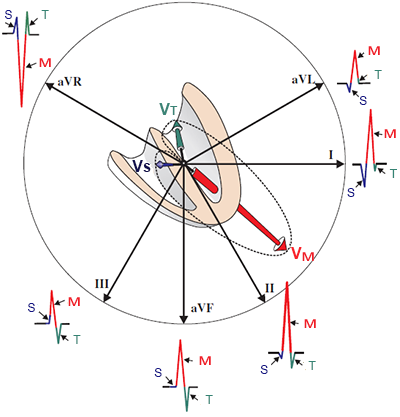
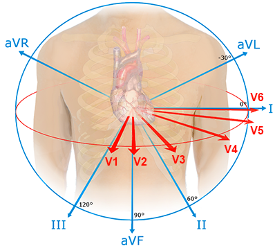
Limb Leads and R Wave
|
|
Pathological R Wave
|
|
ECG and Dominant R Wave in aVR
|
 Ventricular Tachycardia |
Ventricular Tachycardia
Tricyclic Antidepressants (Intoxication)
Dextrocardia
Swapped ECG Leads (Left and Right Arm)
Correctly Placed Limb ECG Leads
Acute Pulmonary Embolism
Sources