ECGbook.com

Making Medical Education Free for All

ECGbook.com

Making Medical Education Free for All

Home /

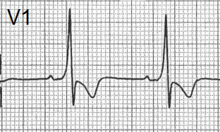
Dominant R Wave in V1

Dominant R wave in V1 ECG lead

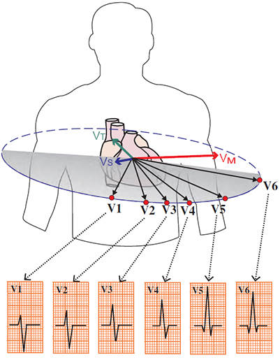
Precordial Leads and R Wave

Dominant R wave V1 in chest leads (V1-V6) explained
  • Precordial leads (V1-V6)
    • view the heart in the horizontal plane

  • Main Cardiac Vector (VM)
    • Points most directly towards lead V6
      • There are lungs (insulator) between the vector and V6
    • Dominant R wave is in V5
    • Points away from lead V1
      • where there will be a negative S wave

Pathological R Wave

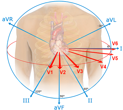
Dominant R wave in V1 and main electrical vector depolarization in ECG leads

ECG and Dominant R Wave in V1

  • Dominant R in V1
    • R wave ≥ S wave
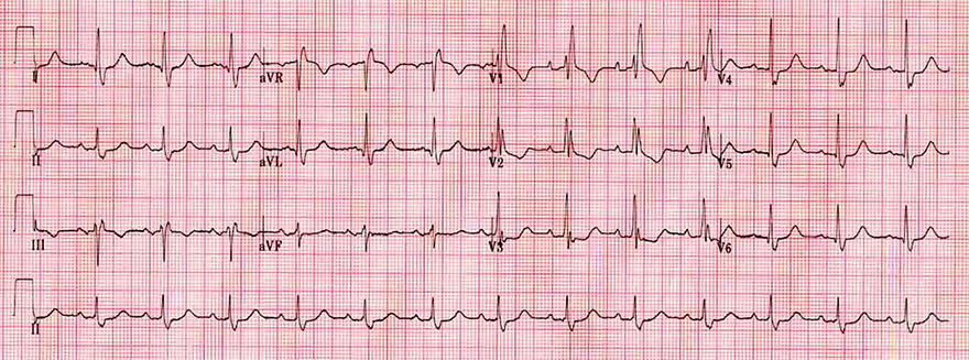
ECG dominant R wave V1 and WPW syndrome type 1, delta wave
WPW Syndrome (Type A)

Causes of Dominant R Wave in V1


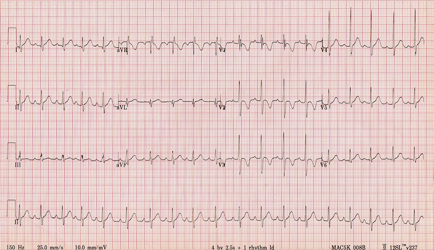
ECG 2 years old child with sinus tachycardi, right ventricular hypertrophy and dominant R wave in V1

Sinus Tachycardia (2-Year-Old Child)

  • 2-Year-Old Healthy Child
    • Children have right ventricular hypertrophy from birth
      • The right ventricle pumps blood against higher pressure in the womb
    • The left ventricle starts to become dominant from the age of 4-6 years
    • Sinus Tachycardia at 110/min is physiological in a 2-year-old child
  • Dominant R in V1
    • It is due to right ventricular hypertrophy
    • It disappears at an older age (4-6 years)


ECG dominant R wave V1 due to right ventricular hypertrophy

Right Ventricular Hypertrophy


ECG dominant R wave V1 and complete right bundle branch block (RBBB)

Right Tawar Bundle Branch Block


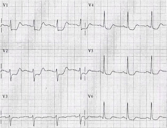
ECG dominant r wave V1 and posterior STEMI infarction

STEMI of the Posterior Wall


ECG dominant r wave V1 and WPW syndrome type A

WPW Syndrome (Type A)

  • WPW Syndrome Type A
    • Shortened PQ interval < 0.12s
    • Delta wave - broadened lower part of the QRS, most visible in (I, II, V4-V6)
    • Type A (Kent bundle is in the left ventricle, resulting in a positive QRS in V1)
  • Dominant R wave in V1
    • Ventricles are activated via 2 pathways (through the AV node and Kent bundle)
    • The ventricular vector then points towards the right ventricle (towards leads V1-V3)


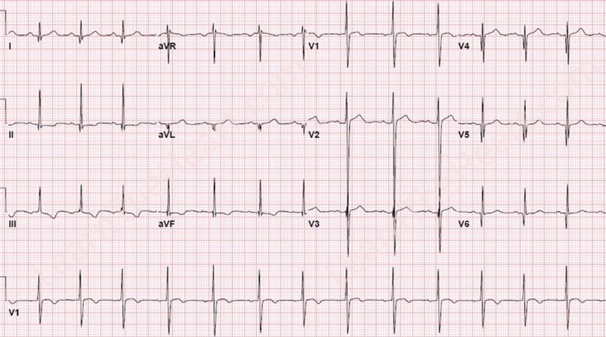
ECG precordial V1 V3 lead misplacement and dominant R wave V1

Swapped Leads V1 and V3


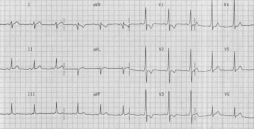
ECG dominant R wave V1 and dextrocardia

Dextrocardia


ECG dominant R wave V1 and hypertrophic cardiomyopathy

Hypertrophic Cardiomyopathy


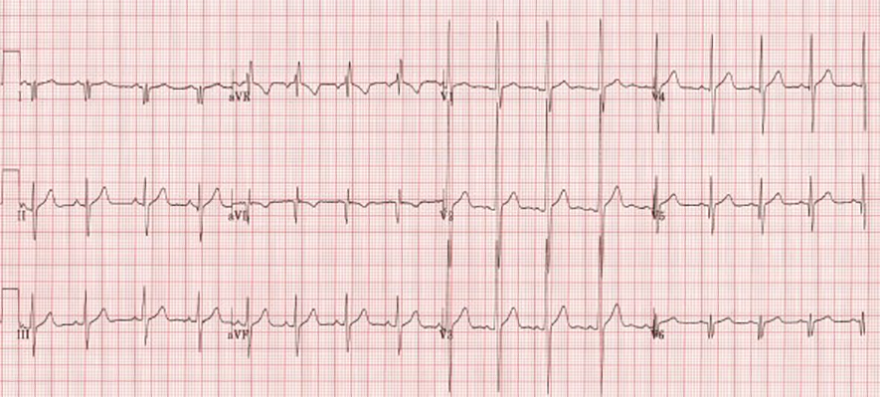
ECG dominant R wave V1 and muscular dystrophy

Muscular Dystrophy

  • Muscular Dystrophy
    • It is a group of inherited disorders characterized by atrophy of muscle tissue throughout the body, including the heart
  • Non-specific changes occur on the EKG
  • The EKG is from a patient with muscular dystrophy
  • Dominant R wave in V1
    • Probably, the left ventricle is more atrophied than the right
    • The vector from the dominant right ventricle points toward leads V1-V3


Sources

  • ECG from Basics to Essentials Step by Step
  • litfl.com
  • ecgwaves.com
  • metealpaslan.com
  • medmastery.com
  • uptodate.com
  • ecgpedia.org
  • wikipedia.org
  • Strong Medicine
  • Understanding Pacemakers


šípka späť

Dominant R Wave in V1

Dominant R wave in V1 ECG lead

Precordial Leads and R Wave

  • Precordial leads (V1-V6)
    • view the heart in the horizontal plane

  • Main Cardiac Vector (VM)
    • Points most directly towards lead V6
      • There are lungs (insulator) between the vector and V6
    • Dominant R wave is in V5
    • Points away from lead V1
      • where there will be a negative S wave

Dominant R wave V1 in chest leads (V1-V6) explained

Pathological R Wave

  • Physiologically, the main vector in 3D space
    • Points from lead aVR (Negative S wave, Q wave)
    • Towards the lateral leads (aVL, I, V5, V) (Dominant R wave)

  • Following R waves require increased attention:
  • Dominant R wave in V1
    • The R wave should not be dominant in lead V1
  • Dominant R wave in aVR
    • The R wave should not be dominant in lead aVR
  • Decreased R wave progression
    • The height of the R wave does not increase from V1 to V5
  • QRS alternans
    • The height of the R wave changes from beat to beat

Dominant R wave in V1 and main electrical vector depolarization in ECG leads

ECG and Dominant R Wave in V1

  • Dominant R in V1
    • R wave ≥ S wave

ECG dominant R wave V1 and WPW syndrome type 1, delta wave
WPW Syndrome (Type A)

Causes of Dominant R Wave in V1


ECG 2 years old child with sinus tachycardi, right ventricular hypertrophy and dominant R wave in V1

Sinus Tachycardia (2-Year-Old Child)

  • 2-Year-Old Healthy Child
    • Children have right ventricular hypertrophy from birth
      • The right ventricle pumps blood against higher pressure in the womb
    • The left ventricle starts to become dominant from the age of 4-6 years
    • Sinus Tachycardia at 110/min is physiological in a 2-year-old child
  • Dominant R in V1
    • It is due to right ventricular hypertrophy
    • It disappears at an older age (4-6 years)


ECG dominant R wave V1 due to right ventricular hypertrophy

Right Ventricular Hypertrophy


ECG dominant R wave V1 and complete right bundle branch block (RBBB)

Right Tawar Bundle Branch Block


ECG dominant r wave V1 and posterior STEMI infarction

STEMI of the Posterior Wall


ECG dominant r wave V1 and WPW syndrome type A

WPW Syndrome (Type A)

  • WPW Syndrome Type A
    • Shortened PQ interval < 0.12s
    • Delta wave - broadened lower part of the QRS, most visible in (I, II, V4-V6)
    • Type A (Kent bundle is in the left ventricle, resulting in a positive QRS in V1)
  • Dominant R wave in V1
    • Ventricles are activated via 2 pathways (through the AV node and Kent bundle)
    • The ventricular vector then points towards the right ventricle (towards leads V1-V3)


ECG precordial V1 V3 lead misplacement and dominant R wave V1

Swapped Leads V1 and V3


ECG dominant R wave V1 and dextrocardia

Dextrocardia


ECG dominant R wave V1 and hypertrophic cardiomyopathy

Hypertrophic Cardiomyopathy


ECG dominant R wave V1 and muscular dystrophy

Muscular Dystrophy

  • Muscular Dystrophy
    • It is a group of inherited disorders characterized by atrophy of muscle tissue throughout the body, including the heart
  • Non-specific changes occur on the EKG
  • The EKG is from a patient with muscular dystrophy
  • Dominant R wave in V1
    • Probably, the left ventricle is more atrophied than the right
    • The vector from the dominant right ventricle points toward leads V1-V3


Sources

  • ECG from Basics to Essentials Step by Step
  • litfl.com
  • ecgwaves.com
  • metealpaslan.com
  • medmastery.com
  • uptodate.com
  • ecgpedia.org
  • wikipedia.org
  • Strong Medicine
  • Understanding Pacemakers