|
|
ECGbook.com Making Medical Education Free for All |
Upload ECG for Interpretation |
|
|
ECGbook.com Making Medical Education Free for All |
Upload ECG for Interpretation |
|
|
ECGbook.com Making Medical Education Free for All |
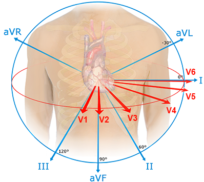

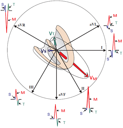
Limb Leads
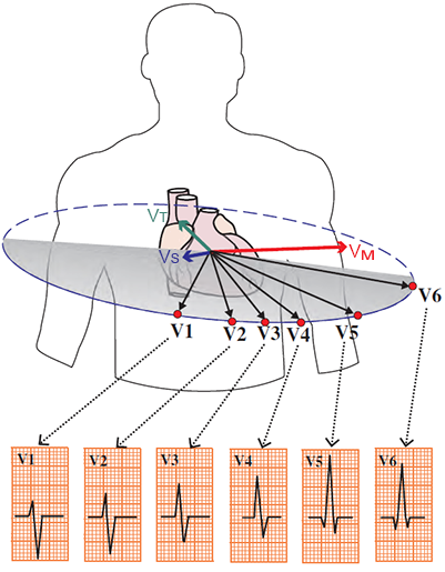
Chest Leads
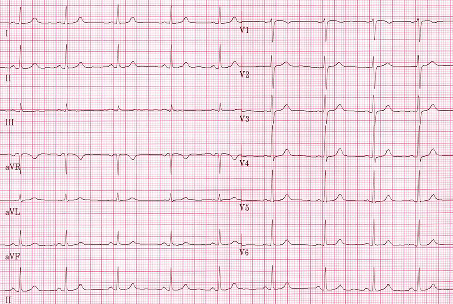
Sinus Rhythm
Sources
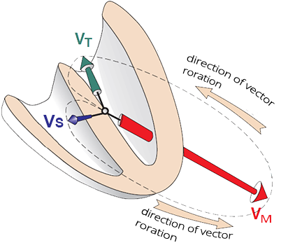
Main Ventricular Vector
|
|
ECG Leads
|
|
|
|
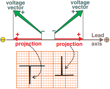
Positive and Negative ECG Deflection
|
|
|
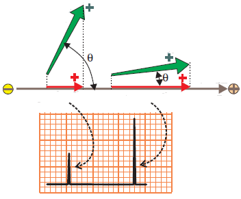
Magnitude of the ECG Deflection
|
|
|
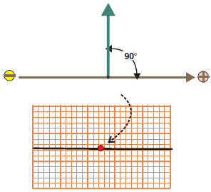
Isoelectric ECG Deflection
|
|
|
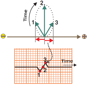
Biphasic ECG Deflection
|
|
|
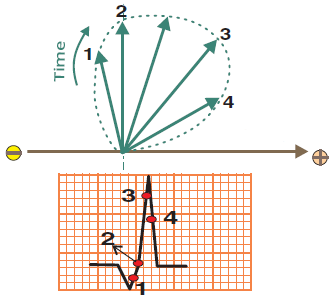
Predominantly Positive ECG Deflection
|
|
|
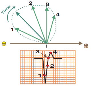
Predominantly Negative ECG Deflection
|
|
|
|
|
Limb Leads
|
Chest Leads
|
Sinus Rhythm
Sources