|
|
ECGbook.com Making Medical Education Free for All |
|
|
ECGbook.com Making Medical Education Free for All |
|
|
ECGbook.com Making Medical Education Free for All |
Home /
ECG patterns of depolarization during ventricular pacing

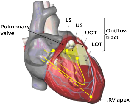

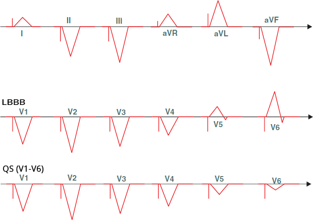
Ventricular Pacing from the Right Ventricular Apex

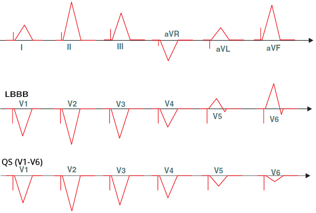
Ventricular Pacing from the Right Ventricular Outflow Tract


Ventricular Pacing from the Left Ventricle

Biventricular Ventricular Pacing
Sources
Home /
ECG patterns of depolarization during ventricular pacing
Ventricular Electrode
|
|
|
|
|
|
Ventricular Pacing from the Right Ventricular Apex
|
|
Ventricular Pacing from the Right Ventricular Outflow Tract
|
|
Ventricular Pacing from the Left Ventricle
Biventricular Ventricular Pacing
Sources