ECGbook.com

Making Medical Education Free for All

ECGbook.com

Making Medical Education Free for All

Home /

ECG Electrodes and Leads

ECG electrodes and ECG leads

ECG Electrodes

Four extremity ecg electrodes and six chest ecg electrodes
  • The ECG device has 10 electrodes that are applied on the body
    • Limb electrodes (4)
      • Attached to the limbs
      • The black limb electrode is the ground
    • Chest electrodes (6)
      • Applied to the chest
  • The electrodes record the electrical vector of the myocardium
  • The ECG device displays the vector as an ECG waveform
  • The ECG paper has 12 leads:
    • V1, V2, V3, V4, V5, V6
    • I, II, III, aVF, aVL, aVR

Limb Electrodes

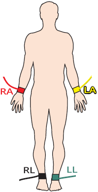
ECG extremity electrodes, left arm (LA), right arm (RA), right leg - neutral, left leg

Chest Electrodes

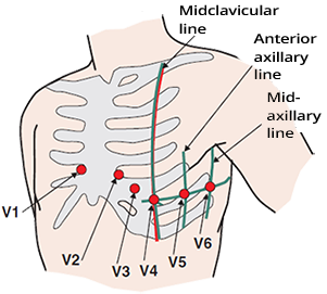
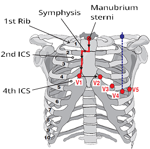
ECG chest electrodes (V1, V2, V3, V4, V5, V6) and precordial position
  • Chest electrodes are suctioned onto the chest
    • Placement is important

  • Electrode placement
    • V1 - 4th intercostal space, right sternal border
    • V2 - 4th intercostal space, left sternal border
    • V3 - Exactly between V2 and V4
    • V4 - 5th intercostal space, midclavicular line
    • V5 - 5th intercostal space, anterior axillary line
    • V6 - 5th intercostal space, midaxillary line

  • Chest electrodes appear on the ECG paper as:
    • Chest leads (V1-V6)

Placement of Chest Electrodes

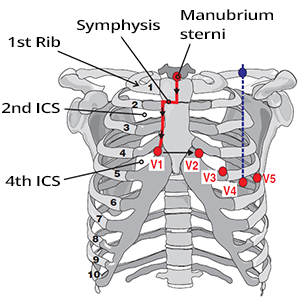
ECG chest electrodes and locating 4th intercostal space

Voltmeter and ECG

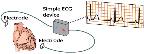
ecg electrodes, voltmeter and ecg paper

Simple ECG Device

  • An ECG device is a voltmeter that records volts as an ECG curve
    • Volts are generated during the propagation of the electrical vector through the myocardium
  • The voltmeter (ECG) always has one electrode positive and the other negative
  • If there is a depolarized (-) and repolarized (+) part of the myocardium between the electrodes (electrical vector)
    • Then the voltmeter measures the electric voltage, which the ECG displays as spikes or waves
  • If there is only a depolarized (-) or only a repolarized (+) part of the myocardium between the electrodes
    • Then the voltmeter does not measure any electric voltage, which the ECG displays as an isoelectric line

3 Limb Electrodes

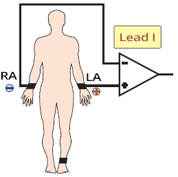
ECG bipolar I lead between two electrodes right arm (RA) and left arm (LA)
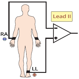
ECG bipolar II lead between two electrodes right arm (RA) and left leg (LL)
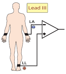
ECG bipolar III lead between two electrodes left arm (LA) and left leg (LL)

Limb Electrodes and Limb Leads


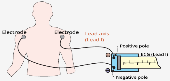
Principle of Lead I

Registration ECG lead I axis with positive and negative pole

Lead I

  • 3 limb electrodes are placed on the wrists and left ankle
  • The body is a good electrical conductor, so it doesn't matter if you place the electrode
    • on the shoulder or on the wrist
    • on the thigh or on the ankle
  • The picture shows Lead I
    • Negative electrode on the right shoulder
    • Positive electrode on the left shoulder

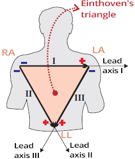
Einthoven's Triangle

Einthoven's equilateral triangle with ECG electrodes and leads

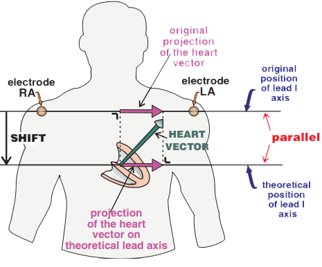
Electrical Axis of ECG Lead

Projection of the heart vector. Original and theoretical lead I axis with parallel shift
  • Each bipolar ECG lead (I, II, III) has
    • a positive (+) and a negative (-) electrode
    • an electrical axis between the (+) and (-) electrodes

  • The heart's electrical vector
    • projects onto the electrical axis of the ECG lead

  • Parallel shifting of the ECG lead (I, II, III)
    • does not change the vector magnitude on the electrical axis

Wilson Central Terminal (WCT)

Wilson central terminal is the hypothetical electrical center of the heart

Einthoven ECG Leads

  • Electrodes are placed on the patient's body (there are 10 of them)
  • On the ECG paper, we see ECG leads (there are 12 of them) generated by the ECG device from the placed electrodes

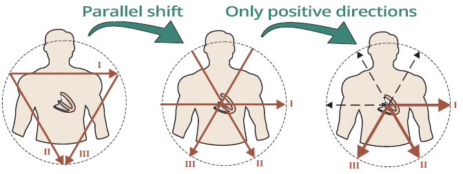
Bipolar ECG leads and parallel shift to Wilson central terminal

Einthoven ECG Leads

  • Leads (I, II, III) can be shifted in parallel towards the central terminal
    • The Wilson central terminal serves as a negative electrode calculated by the ECG device
  • The electrical axis of each lead (I, II, III)
    • directs from the terminal towards the positive electrode

Goldberger ECG Leads

Goldberger augmented leads (aVR, aVL, aVF)

Hexaxial System

  • From 3 electrodes placed on the limbs
  • The ECG device generates 6 ECG leads
    • Einthoven leads (I, II, III)
    • Goldberger leads (aVR, aVF, aVL)
  • The leads (I, II, III, aVR, aVF, aVL) together are referred to as limb leads

ECG hexaxial diagram with einthoven leads (I, II, III) and goldberger leads (aVR, aVF, aVL)

Hexaxial System

  • Created by combining Einthoven and Goldberger leads
    • From central terminal, 6 ECG leads are oriented
  • Each ECG lead "views" the heart from a different angle of perspective
    • These 6 ECG leads are sometimes referred to as the hexaxial system
  • Limb leads "view" the heart in the frontal plane

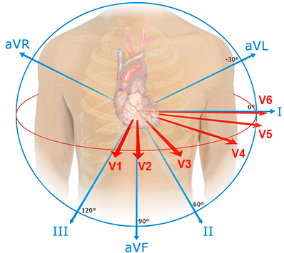
Chest Electrodes

Wilson unipolar chest precordial leads (V1, V2, V3, V4, V5, V6)

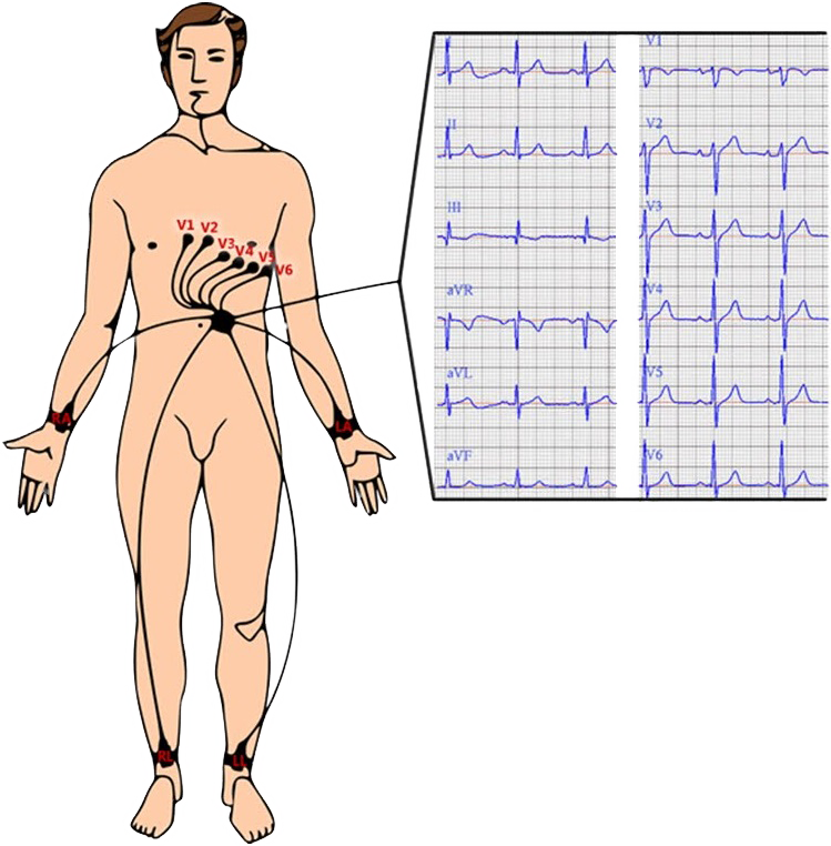
12-Lead ECG

  • The ECG device has 10 electrodes that are attached to the body
    • The black electrode serves as ground and does not contribute to generating the ECG waveform
    • Thus, the ECG device uses 9 electrodes for generating the ECG waveform

  • The ECG paper displays 12 leads, which the ECG device generates from the attached electrodes

  • Standard limb leads (I, II, III, aVR, aVF, aVL):
    • Einthoven's leads (I, II, III)
      • These are bipolar because they are derived from a positive and a negative electrode
    • Goldberger's leads (aVR, aVF, aVL)
      • These are unipolar because they are derived only from a positive electrode (and the central terminal)

  • Wilson's chest leads (V1-V6)
    • These are unipolar because they are derived only from chest electrodes (and the central terminal)

ECG chest and limb electrodes position and ECG leads

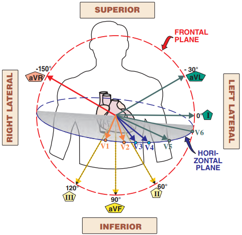
ECG Leads in 3D Space

12 ECG leads in 3D space and main heart vector and Wilson central terminal

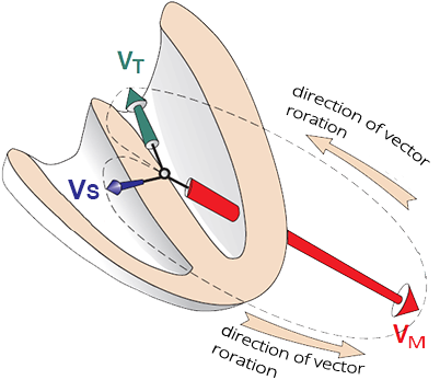
Ventricular Vectors

Summary of the frontal plane heart vectors directions
  • The thin ventricular septum is first activated
    • Resulting in a small septal vector (VS)
    • Q wave
  • Then the massive left ventricle is activated
    • Resulting in a large main vector (VM)
    • Simultaneously, the thin right ventricle is also activated
      • Small right ventricular vector
      • does not influence the direction of the main vector
    • R wave
  • Finally, the base of the left ventricle is activated
    • Resulting in a small terminal vector (VT)
    • S wave

  • You must imagine the vectors in 3D space
    • ECG leads record vectors over time

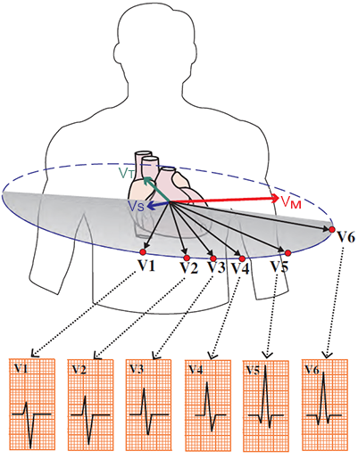
ECG and Ventricular Vectors

Limb leads: Initial vector (Q wave), main vector (R wave), terminal vector (S wave)

Limb Leads


Chest leads: Initial vector (Q wave), main vector (R wave), terminal vector (S wave)

Chest Leads


12 leads ECG with sinus rhythm

Sinus Rhythm

  • On the ECG is a sinus rhythm
  • Note how the height of the QRS complexes changes in the different leads

ECG Leads and 12-Lead ECG

ECG leads in 3D space (superior, right lateral, inferior, left lateral leads)

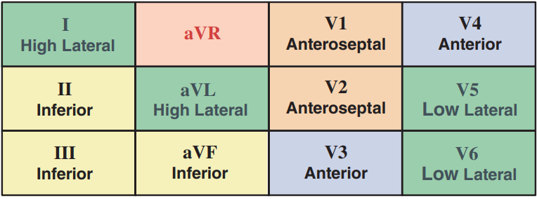
ECG paper leads position: high lateral (I, aVL), inferior (II, III, aVF), anteroseptal (V1, V2), anterior (V3, V4), low lateral (V5, V6), forgotten aVR lead

12-Lead ECG


Chest Leads (V1-V2)

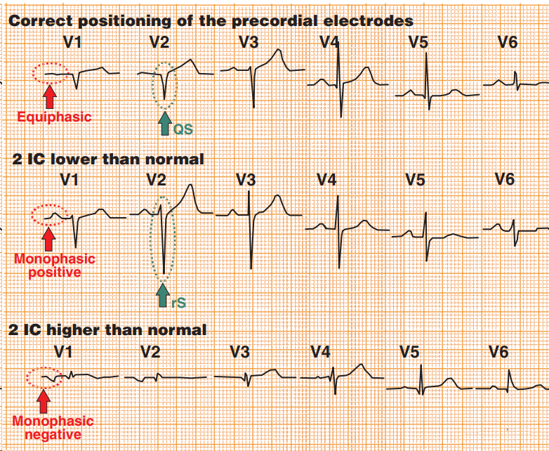
Correct position of the precordial electrodes
Correct and incorrect position V1, V2. And differential diagnosis between left anterior hemiblock (LAH) and old anteroseptal infarction

Differential Diagnosis with V1-V2

  • Correct placement of electrodes V1-V2 in the 4th intercostal space
  • If V1-V2 is placed lower (6th intercostal space)
    • P wave is monophasic positive
    • rS configuration (V1-V2) appears
      • This indicates a Left Anterior Hemiblock
      • Old infarction would still show a QS configuration (without r wave)
  • If V1-V2 is placed higher (2nd intercostal space)
    • P wave is monophasic negative


Sources

  • ECG from Basics to Essentials Step by Step
  • litfl.com
  • ecgwaves.com
  • metealpaslan.com
  • medmastery.com
  • uptodate.com
  • ecgpedia.org
  • wikipedia.org
  • Strong Medicine
  • Understanding Pacemakers


šípka späť

ECG Electrodes and Leads

ECG electrodes and ECG leads

ECG Electrodes

  • The ECG device has 10 electrodes that are applied on the body
    • Limb electrodes (4)
      • Attached to the limbs
      • The black limb electrode is the ground
    • Chest electrodes (6)
      • Applied to the chest
  • The electrodes record the electrical vector of the myocardium
  • The ECG device displays the vector as an ECG waveform
  • The ECG paper has 12 leads:
    • V1, V2, V3, V4, V5, V6
    • I, II, III, aVF, aVL, aVR
Four extremity ecg electrodes and six chest ecg electrodes

Limb Electrodes

  • Limb electrodes are clipped onto the limbs
    • on the wrists and ankles
    • Height does not matter

  • Electrode placement
    • Red - right arm (RA)
    • Yellow - left arm (LA)
    • Green - left leg (LL)
    • Black - right leg (RL)
  • Using 3 limb electrodes
    • The ECG machine generates 6 ECG leads on the paper:
      • Limb leads (I, II, III, aVL, aVF, aVR)

ECG extremity electrodes, left arm (LA), right arm (RA), right leg - neutral, left leg

Chest Electrodes

  • Chest electrodes are suctioned onto the chest
    • Placement is important

  • Electrode placement
    • V1 - 4th intercostal space, right sternal border
    • V2 - 4th intercostal space, left sternal border
    • V3 - Exactly between V2 and V4
    • V4 - 5th intercostal space, midclavicular line
    • V5 - 5th intercostal space, anterior axillary line
    • V6 - 5th intercostal space, midaxillary line

  • Chest electrodes appear on the ECG paper as:
    • Chest leads (V1-V6)

ECG chest electrodes (V1, V2, V3, V4, V5, V6) and precordial position

Placement of Chest Electrodes

  • Key is locating the 4th intercostal space (ICS)
  • Feel for the upper part of the sternum (manubrium sterni)
  • Move down to the palpable notch (manubrio-sternal symphysis)
    • Then move right parasternally (to the edge of the sternum)
    • You will reach the 2nd rib
  • Move down parasternally and count the intercostal spaces
  • You will reach the 4th intercostal space (V1)
    • Move left parasternally (V2)
  • Feel for the 5th intercostal space and midclavicular line (V4)
  • Lead V3 is placed exactly between V2 and V4
  • Leads V5-V6 are at the same level as V4
    • V5 (anterior axillary line)
    • V6 (midaxillary line)
ECG chest electrodes and locating 4th intercostal space

Voltmeter and ECG

ecg electrodes, voltmeter and ecg paper

Simple ECG Device

  • An ECG device is a voltmeter that records volts as an ECG curve
    • Volts are generated during the propagation of the electrical vector through the myocardium
  • The voltmeter (ECG) always has one electrode positive and the other negative
  • If there is a depolarized (-) and repolarized (+) part of the myocardium between the electrodes (electrical vector)
    • Then the voltmeter measures the electric voltage, which the ECG displays as spikes or waves
  • If there is only a depolarized (-) or only a repolarized (+) part of the myocardium between the electrodes
    • Then the voltmeter does not measure any electric voltage, which the ECG displays as an isoelectric line

3 Limb Electrodes

ECG bipolar I lead between two electrodes right arm (RA) and left arm (LA) ECG bipolar II lead between two electrodes right arm (RA) and left leg (LL) ECG bipolar III lead between two electrodes left arm (LA) and left leg (LL)

Limb Electrodes and Limb Leads


Principle of Lead I

Registration ECG lead I axis with positive and negative pole

Lead I

  • 3 limb electrodes are placed on the wrists and left ankle
  • The body is a good electrical conductor, so it doesn't matter if you place the electrode
    • on the shoulder or on the wrist
    • on the thigh or on the ankle
  • The picture shows Lead I
    • Negative electrode on the right shoulder
    • Positive electrode on the left shoulder

Einthoven's Triangle

Einthoven's equilateral triangle with ECG electrodes and leads
  • 3 limb electrodes (right arm, left arm, left thigh)
    • form the Einthoven's triangle

  • This creates 3 bipolar ECG leads:
  • bipolar because each ECG lead has 2 electrodes
    • Lead I: right arm (-) and left arm (+)
    • Lead II: right arm (-) and left thigh (+)
    • Lead III: left arm (+) and left thigh (-)

Electrical Axis of ECG Lead

Projection of the heart vector. Original and theoretical lead I axis with parallel shift
  • Each bipolar ECG lead (I, II, III) has
    • a positive (+) and a negative (-) electrode
    • an electrical axis between the (+) and (-) electrodes

  • The heart's electrical vector
    • projects onto the electrical axis of the ECG lead

  • Parallel shifting of the ECG lead (I, II, III)
    • does not change the vector magnitude on the electrical axis

Wilson Central Terminal (WCT)

Wilson central terminal is the hypothetical electrical center of the heart
  • Each ECG lead must have
    • a positive and a negative electrode
    • between which there is an electrical axis of the lead

  • Einthoven ECG leads (I, II, III)
    • are bipolar
      (having a positive and negative electrode)

  • Wilson connected 3 limb electrodes to resistors
    • and created an electrical center of the heart
    • it serves as a reference negative electrode
    • known as Wilson central terminal (WCT)
  • The ECG device based on 3 limb electrodes
    • creates a reference electrode at the center of the triangle

  • The Wilson central terminal is located:

Einthoven ECG Leads

  • Electrodes are placed on the patient's body (there are 10 of them)
  • On the ECG paper, we see ECG leads (there are 12 of them) generated by the ECG device from the placed electrodes

Bipolar ECG leads and parallel shift to Wilson central terminal

Einthoven ECG Leads

  • Leads (I, II, III) can be shifted in parallel towards the central terminal
    • The Wilson central terminal serves as a negative electrode calculated by the ECG device
  • The electrical axis of each lead (I, II, III)
    • directs from the terminal towards the positive electrode

Goldberger ECG Leads

Goldberger augmented leads (aVR, aVL, aVF)
  • The ECG device generates from 3 electrodes:
    • Einthoven leads (I, II, III)
    • Goldberger leads (aVR, aVF, aVL)

  • Goldberger leads (aVR, aVF, aVL) are oriented
    • From the central terminal towards the limb electrodes

  • Goldberger leads are augmented (extended)
    • If the ECG device measured them from the terminal to the electrodes (+)
      • the ECG curve would be very low
    • The ECG device measures them
      • from the center of the opposite side of Einthoven's triangle
      • The ECG signal is thus 50% higher
        • thus they are called augmented (extended)

  • aVR (augmented vector right)
  • aVL (augmented vector left)
  • aVF (augmented vector foot)

Hexaxial System

  • From 3 electrodes placed on the limbs
  • The ECG device generates 6 ECG leads
    • Einthoven leads (I, II, III)
    • Goldberger leads (aVR, aVF, aVL)
  • The leads (I, II, III, aVR, aVF, aVL) together are referred to as limb leads

ECG hexaxial diagram with einthoven leads (I, II, III) and goldberger leads (aVR, aVF, aVL)

Hexaxial System

  • Created by combining Einthoven and Goldberger leads
    • From central terminal, 6 ECG leads are oriented
  • Each ECG lead "views" the heart from a different angle of perspective
    • These 6 ECG leads are sometimes referred to as the hexaxial system
  • Limb leads "view" the heart in the frontal plane

Chest Electrodes

  • Applied to the chest
  • The electrical axis of the chest leads points
    • from the central terminal
    • to each applied chest electrode
  • "View" the heart in the horizontal plane
  • Sometimes referred to as Wilson's EKG leads

Wilson unipolar chest precordial leads (V1, V2, V3, V4, V5, V6)

12-Lead ECG

  • The ECG device has 10 electrodes that are attached to the body
    • The black electrode serves as ground and does not contribute to generating the ECG waveform
    • Thus, the ECG device uses 9 electrodes for generating the ECG waveform

  • The ECG paper displays 12 leads, which the ECG device generates from the attached electrodes

  • Standard limb leads (I, II, III, aVR, aVF, aVL):
    • Einthoven's leads (I, II, III)
      • These are bipolar because they are derived from a positive and a negative electrode
    • Goldberger's leads (aVR, aVF, aVL)
      • These are unipolar because they are derived only from a positive electrode (and the central terminal)

  • Wilson's chest leads (V1-V6)
    • These are unipolar because they are derived only from chest electrodes (and the central terminal)

ECG chest and limb electrodes position and ECG leads

ECG Leads in 3D Space

  • 12 ECG leads "view" the heart in 2 planes:
    • Horizontal plane (V1-V6)
    • Frontal plane (I, II, III, aVF, aVR, aVL)
  • All leads originate from the central terminal
  • If the electrical vector points
    • Towards the lead - a positive deflection is produced
    • Away from the lead - a negative deflection is produced


12 ECG leads in 3D space and main heart vector and Wilson central terminal

Ventricular Vectors

  • The thin ventricular septum is first activated
    • Resulting in a small septal vector (VS)
    • Q wave
  • Then the massive left ventricle is activated
    • Resulting in a large main vector (VM)
    • Simultaneously, the thin right ventricle is also activated
      • Small right ventricular vector
      • does not influence the direction of the main vector
    • R wave
  • Finally, the base of the left ventricle is activated
    • Resulting in a small terminal vector (VT)
    • S wave

  • You must imagine the vectors in 3D space
    • ECG leads record vectors over time

Summary of the frontal plane heart vectors directions

ECG and Ventricular Vectors

Limb leads: Initial vector (Q wave), main vector (R wave), terminal vector (S wave) Chest leads: Initial vector (Q wave), main vector (R wave), terminal vector (S wave)

Limb Leads

Chest Leads


12 leads ECG with sinus rhythm

Sinus Rhythm

  • On the ECG is a sinus rhythm
  • Note how the height of the QRS complexes changes in the different leads

ECG Leads and 12-Lead ECG

ECG leads in 3D space (superior, right lateral, inferior, left lateral leads)

ECG paper leads position: high lateral (I, aVL), inferior (II, III, aVF), anteroseptal (V1, V2), anterior (V3, V4), low lateral (V5, V6), forgotten aVR lead

12-Lead ECG


Chest Leads (V1-V2)

Correct and incorrect position V1, V2. And differential diagnosis between left anterior hemiblock (LAH) and old anteroseptal infarction

Differential Diagnosis with V1-V2

  • Correct placement of electrodes V1-V2 in the 4th intercostal space
  • If V1-V2 is placed lower (6th intercostal space)
    • P wave is monophasic positive
    • rS configuration (V1-V2) appears
      • This indicates a Left Anterior Hemiblock
      • Old infarction would still show a QS configuration (without r wave)
  • If V1-V2 is placed higher (2nd intercostal space)
    • P wave is monophasic negative
Correct position of the precordial electrodes


Sources

  • ECG from Basics to Essentials Step by Step
  • litfl.com
  • ecgwaves.com
  • metealpaslan.com
  • medmastery.com
  • uptodate.com
  • ecgpedia.org
  • wikipedia.org
  • Strong Medicine
  • Understanding Pacemakers