ECGbook.com

Making Medical Education Free for All

ECGbook.com

Making Medical Education Free for All

Home /

Focal Atrial Tachycardia

Focal atrial tachycardia, Paroxysmal atrial tachycardia (PAT), Unifocal atrial tachycardia, Ectopic atrial tachycardia
Ectopic Focus

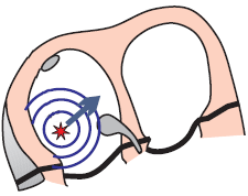
Ectopic atrial focus
  • Most often occurs in a structurally altered atrium
    • Never located in the SA node
  • Has a size of up to 5mm
  • Generates impulses at a frequency of 130-250/min.
  • The focus can start generating impulses through 3 mechanisms:
    • Increased automaticity
    • Triggering activity
    • Micro-reentry
  • Focal atrial tachycardia with micro-reentry is referred to as

Focus Localization

atrial ectopic focus, right atrial, left atrial

Physiological P Wave

Normal P wave electrical vector, positive P wave II, Inverted P wave aVR lead

ECG and Focal Atrial Tachycardia


ECG Unifocal atrial tachycardia, abnormal P wave morphology

Focal Atrial Tachycardia


Differential Diagnosis


atrial ectopic focus, enhanced automaticity, focal atrial tachycardia
ECG focal atrial tachycardia, enhanced automaticity, abnormal P wave

Focal Atrial Tachycardia


atrial reentry, Intra-atrial reentry/reentrant tachycardia (IART)
ECG Intra-atrial reentry/reentrant tachycardia (IART)

Intra-Atrial Reentry Tachycardia

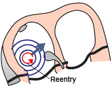
  • Intra-atrial reentry tachycardia
    • It is a focal atrial tachycardia with a micro-reentry mechanism
  • In the atrium, there is an ectopic focus generating impulses instead of the SA node
  • Frequency 100-250/min.
  • P waves have a different shape compared to sinus rhythm
    • In this case, the P waves are negative in lead II
  • Electro-mechanism of the ectopic focus is:
    • Micro-reentry (tachycardia is paroxysmal)


multiple ectopic foci within the atria
ECG Multifocal Atrial Tachycardia (MAT), Chaotic atrial tachycardia

Multifocal Atrial Tachycardia


AV Conduction

carotid sinus massage, unifocal atrial tachycardia

ECG supraventricular tachycardia, 300bpm, narrow QRS

Supraventricular Tachycardia


carotid sinus massage, unifocal atrial tachycardia
ECG AV block, carotid sinus massage

Carotid Sinus Massage


ECG narrow complex supraventricular tachycardia

Supraventricular Tachycardia

  • Frequency: 140/min.
  • One P wave is in the terminal portion of the QRS complex
  • Is the second P wave hidden in the T wave?


carotid sinus massage, unifocal atrial tachycardia
ECG carotid sinus massage, uni-focal atrial tachycardia, abnormal P waves morphology

Carotid Sinus Massage


ECG unifocal atrial tachycardia, fixed AV block 2:1

Focal Atrial Tachycardia and Second-Degree AV Block (2:1)


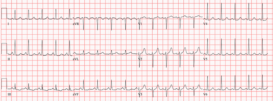
ECG atrial tachycardia, abnormal P waves, narrow QRS

Focal Atrial Tachycardia

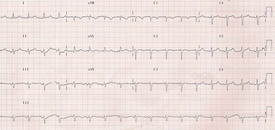
  • Frequency: 120/min.
  • P wave is negative (II, III, aVF) and positive (V1)
    • A physiological P wave is never negative in (II, III, aVF) and positive in (V1)


ECG ectopic atrial tachycardia

Focal Atrial Tachycardia


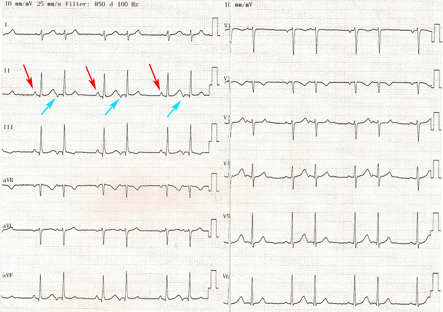
ECG ectopic atrial rhythm, sinus rhythm, abnormal and normal P waves

Focal Atrial Rhythm and Sinus Rhythm

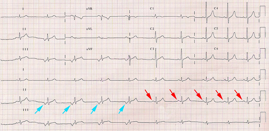
  • This is a healthy 14-year-old boy
  • Focal Atrial Rhythm (blue arrows)
  • Sinus Rhythm follows after 4 negative P waves (red arrows)
    • P wave is now positive in lead II
    • Frequency: 75/min. The SA node increased the frequency and overtook the ectopic focus (overdrive suppression)
  • The focal site does not necessarily generate impulses only with tachycardia frequency (>100/min.)


ECG sinus tachycardia vs. Paroxysmal atrial tachycardia (PAT)

Sinus Tachycardia


ECG atrial bigeminy rhythm, ectopic focus and SA node depolarisation

Atrial Bigeminy Rhythm

  • Sinus Rhythm
    • P waves are positive (II, III, aVF) and negative (V1) (red arrows)
  • Focal Atrial Rhythm
    • P waves are negative (II, III, aVF) and positive (V1) (blue arrows)
    • Essentially, it is regular atrial extrasystoles
  • If sinus rhythm alternates with atrial extrasystoles in a 1:1 ratio


Sources

  • ECG from Basics to Essentials Step by Step
  • litfl.com
  • ecgwaves.com
  • metealpaslan.com
  • medmastery.com
  • uptodate.com
  • ecgpedia.org
  • wikipedia.org
  • Strong Medicine
  • Understanding Pacemakers


šípka späť

Focal Atrial Tachycardia

Focal atrial tachycardia, Paroxysmal atrial tachycardia (PAT), Unifocal atrial tachycardia, Ectopic atrial tachycardia
Ectopic Focus

  • Most often occurs in a structurally altered atrium
    • Never located in the SA node
  • Has a size of up to 5mm
  • Generates impulses at a frequency of 130-250/min.
  • The focus can start generating impulses through 3 mechanisms:
    • Increased automaticity
    • Triggering activity
    • Micro-reentry
  • Focal atrial tachycardia with micro-reentry is referred to as


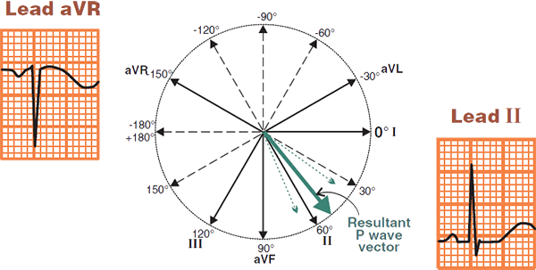
Ectopic atrial focus

Focus Localization

  • The focus occurs in structurally altered atria
    • at anatomically uneven sites
  • Atrial changes are most often caused by:
  • The right atrium is more complex than the left
    • Therefore, the focus more often occurs in the right atrium
    • Focus occurrence:
      • Right atrium (67%)
      • Left atrium (33%)
atrial ectopic focus, right atrial, left atrial

Physiological P Wave


Normal P wave electrical vector, positive P wave II, Inverted P wave aVR lead

ECG and Focal Atrial Tachycardia


ECG Unifocal atrial tachycardia, abnormal P wave morphology

Focal Atrial Tachycardia


Differential Diagnosis


atrial ectopic focus, enhanced automaticity, focal atrial tachycardia ECG focal atrial tachycardia, enhanced automaticity, abnormal P wave

Focal Atrial Tachycardia

  • In the atrium, there is an ectopic focus generating impulses instead of the SA node
  • Frequency 100-250/min.
  • P waves have a different shape compared to sinus rhythm
    • In this case, the P waves are negative in lead II
  • Electro-mechanism of the ectopic focus can be:
    • Increased automaticity (tachycardia "warms up")
    • Triggering activity (tachycardia is paroxysmal)


atrial reentry, Intra-atrial reentry/reentrant tachycardia (IART) ECG Intra-atrial reentry/reentrant tachycardia (IART)

Intra-Atrial Reentry Tachycardia

  • Intra-atrial reentry tachycardia
    • It is a focal atrial tachycardia with a micro-reentry mechanism
  • In the atrium, there is an ectopic focus generating impulses instead of the SA node
  • Frequency 100-250/min.
  • P waves have a different shape compared to sinus rhythm
    • In this case, the P waves are negative in lead II
  • Electro-mechanism of the ectopic focus is:
    • Micro-reentry (tachycardia is paroxysmal)


multiple ectopic foci within the atria ECG Multifocal Atrial Tachycardia (MAT), Chaotic atrial tachycardia

Multifocal Atrial Tachycardia

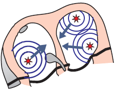
  • Multifocal atrial tachycardia
  • There are at least 3 ectopic foci in the atria
    • Which generate impulses independently of each other
  • Each focus creates a different P wave
    • There are at least 3 different P waves present
  • Frequency 100-250/min.
  • Electro-mechanism of the ectopic foci is most commonly
    • Increased automaticity (tachycardia "warms up")

AV Conduction


carotid sinus massage, unifocal atrial tachycardia


ECG supraventricular tachycardia, 300bpm, narrow QRS

Supraventricular Tachycardia


ECG AV block, carotid sinus massage

Carotid Sinus Massage

  • Carotid sinus massage slows down conduction through the AV node
  • P waves became apparent on the ECG (every 2nd P wave is followed by a QRS)
  • This is Focal Atrial Tachycardia
    • P wave frequency: 300/min
    • QRS complex frequency: 150/min.
  • It could also be atrial flutter
    • Atrial flutter would show typical sawtooth waves in the inferior leads (II, III, aVF)
carotid sinus massage, unifocal atrial tachycardia


ECG narrow complex supraventricular tachycardia

Supraventricular Tachycardia

  • Frequency: 140/min.
  • One P wave is in the terminal portion of the QRS complex
  • Is the second P wave hidden in the T wave?


ECG carotid sinus massage, uni-focal atrial tachycardia, abnormal P waves morphology

Carotid Sinus Massage

carotid sinus massage, unifocal atrial tachycardia


ECG unifocal atrial tachycardia, fixed AV block 2:1

Focal Atrial Tachycardia and Second-Degree AV Block (2:1)


ECG atrial tachycardia, abnormal P waves, narrow QRS

Focal Atrial Tachycardia

  • Frequency: 120/min.
  • P wave is negative (II, III, aVF) and positive (V1)
    • A physiological P wave is never negative in (II, III, aVF) and positive in (V1)


ECG ectopic atrial tachycardia

Focal Atrial Tachycardia


ECG ectopic atrial rhythm, sinus rhythm, abnormal and normal P waves

Focal Atrial Rhythm and Sinus Rhythm

  • This is a healthy 14-year-old boy
  • Focal Atrial Rhythm (blue arrows)
  • Sinus Rhythm follows after 4 negative P waves (red arrows)
    • P wave is now positive in lead II
    • Frequency: 75/min. The SA node increased the frequency and overtook the ectopic focus (overdrive suppression)
  • The focal site does not necessarily generate impulses only with tachycardia frequency (>100/min.)


ECG sinus tachycardia vs. Paroxysmal atrial tachycardia (PAT)

Sinus Tachycardia


ECG atrial bigeminy rhythm, ectopic focus and SA node depolarisation

Atrial Bigeminy Rhythm

  • Sinus Rhythm
    • P waves are positive (II, III, aVF) and negative (V1) (red arrows)
  • Focal Atrial Rhythm
    • P waves are negative (II, III, aVF) and positive (V1) (blue arrows)
    • Essentially, it is regular atrial extrasystoles
  • If sinus rhythm alternates with atrial extrasystoles in a 1:1 ratio


Sources

  • ECG from Basics to Essentials Step by Step
  • litfl.com
  • ecgwaves.com
  • metealpaslan.com
  • medmastery.com
  • uptodate.com
  • ecgpedia.org
  • wikipedia.org
  • Strong Medicine
  • Understanding Pacemakers