|
|
ECGbook.com Making Medical Education Free for All |
|
|
ECGbook.com Making Medical Education Free for All |
|
|
ECGbook.com Making Medical Education Free for All |
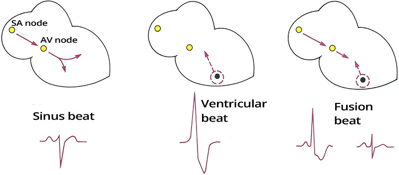
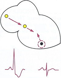
Fusion Beat Mechanism
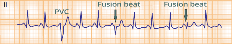
Fusion Beats
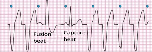
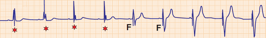
Ventricular Tachycardia
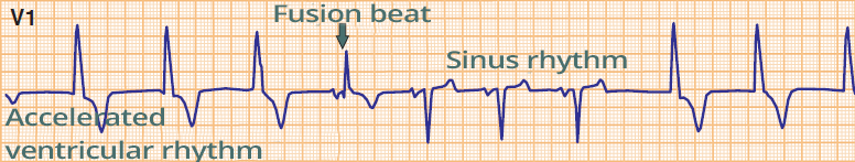
Accelerated Ventricular Rhythm
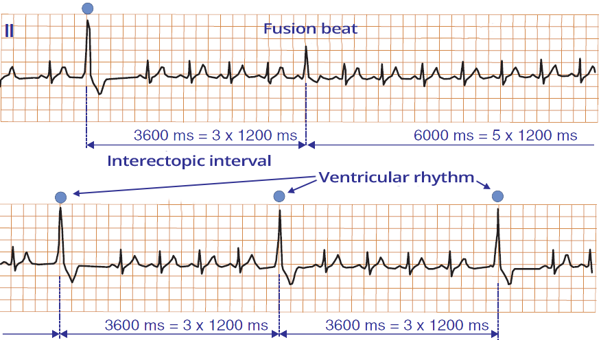
Ventricular Parasystole
Fusion Beat and Pacemaker
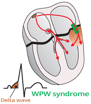
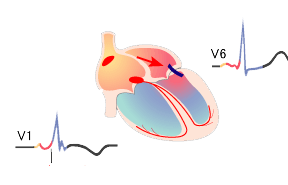
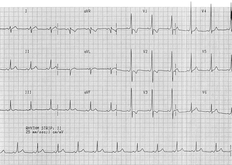
WPW Syndrome (Type A)
Sources
Fusion Beat Mechanism
Fusion Beat
|
|
Fusion Beats
Ventricular Tachycardia
Accelerated Ventricular Rhythm
Ventricular Parasystole
Fusion Beat and Pacemaker
Fusion Beat and WPW Syndrome
|
|
|
WPW Syndrome (Type A)
|
|
Sources