ECGbook.com

Making Medical Education Free for All

ECGbook.com

Making Medical Education Free for All

Home /

Heart and ECG

Heart and ECG

Heart and Circulatory System

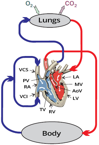
Heart with systemic and pulmonary circulation
  • The heart is a pump that synchronously pumps blood into 2 circulations
    • Into the small circulation (to the lungs)
    • Into the large circulation (to the systemic circulation)
  • The heart as a pump has 2 parts:
  • Into the right atrium (RA)
    • Deoxygenated blood flows in from the large circulation through
      • Vena cava superior (VCS)
      • Vena cava inferior (VCI)
      • and from the heart through the coronary sinus
    • Then it continues through the tricuspid valve (TV)
      • into the right ventricle
  • Right ventricle (RV)
    • Pumps blood to the lungs through the pulmonary valve (PV)

  • Into the left atrium (LA)
    • Oxygenated blood flows in from the lungs
    • Then it continues through the mitral valve (MV)
      • into the left ventricle
  • Left ventricle (LV)
    • Pumps blood to the large circulation through the aortic valve (AoV)

Cardiac Conduction System

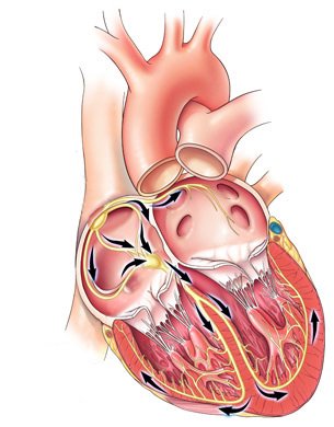
Electrical conduction system of the heart

ECG and Electrical Impulse

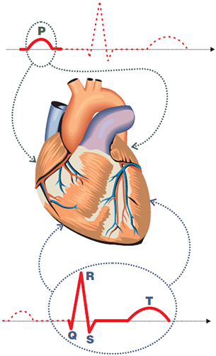
Heart with electrical vectors, with P wave and QRS complex
  • ECG curve represents the spread of impulses through the heart
  • An impulse spreads
  • The ECG curve mainly shows:
    • The slow spread of the impulse in the working myocardium
    • The impulse creates a cardiac vector in the working myocardium

  • Electrical activity in the atrial myocardium creates:
    • The P wave (atrial depolarization)
    • The Ta wave (atrial repolarization)
      • which is hidden in the QRS complex

  • Electrical activity in the ventricular myocardium creates
    • The QRS complex (ventricular depolarization)
    • The T wave (ventricular repolarization)

Main Heart Vector

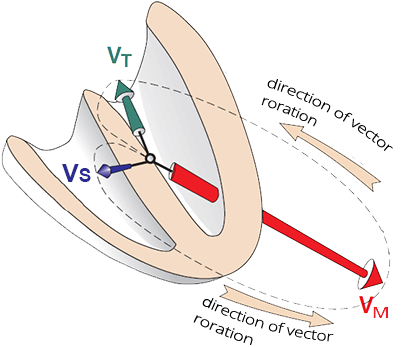
Heart with septal, main, and terminal electrical vector

ECG Leads

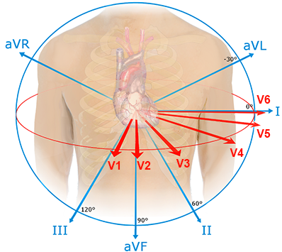
Heart vectors with chest and limb leads
  • ECG recording has 12 leads
  • Each lead "views" the vectors from a different angle
  • The principle of the ECG curve is:
    • If the vector points
      • Toward the lead - a positive wave is recorded
      • Away from the lead - a negative wave is recorded

  • You need to imagine vectors and leads in a 3D space
    • Leads record vectors over time

ECG and the Cardiac Vector

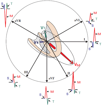
Limb leads with heart vectors, and QRS complex formation

Limb Leads


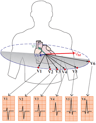
Chest leads with heart vectors, and QRS complex formation

Chest Leads


ECG strip with sinus rhythm

Sinus Rhythm

  • On the ECG, sinus rhythm is present
  • Notice how the height of the QRS complexes changes across different leads

What Does an ECG Diagnose?


Sources

  • ECG from Basics to Essentials Step by Step
  • litfl.com
  • ecgwaves.com
  • metealpaslan.com
  • medmastery.com
  • uptodate.com
  • ecgpedia.org
  • wikipedia.org
  • Strong Medicine
  • Understanding Pacemakers


šípka späť

Heart and ECG

Heart and ECG

Heart and Circulatory System

  • The heart is a pump that synchronously pumps blood into 2 circulations
    • Into the small circulation (to the lungs)
    • Into the large circulation (to the systemic circulation)
  • The heart as a pump has 2 parts:
  • Into the right atrium (RA)
    • Deoxygenated blood flows in from the large circulation through
      • Vena cava superior (VCS)
      • Vena cava inferior (VCI)
      • and from the heart through the coronary sinus
    • Then it continues through the tricuspid valve (TV)
      • into the right ventricle
  • Right ventricle (RV)
    • Pumps blood to the lungs through the pulmonary valve (PV)

  • Into the left atrium (LA)
    • Oxygenated blood flows in from the lungs
    • Then it continues through the mitral valve (MV)
      • into the left ventricle
  • Left ventricle (LV)
    • Pumps blood to the large circulation through the aortic valve (AoV)
Heart with systemic and pulmonary circulation

Cardiac Conduction System

  • The heart has 2 basic functional parts:
    • Conduction system
      • They are thin "wires" that conduct impulses throughout the heart
      • Impulses spontaneously originate in the SA node
        • they spread through the heart and activate the working myocardium
    • Working myocardium
      • It is the heart muscle, which contracts upon stimulation by an impulse
      • This results in atrial systole, followed by ventricular systole

  • ECG device has electrodes that are attached to the body
    • ECG detects the spread of electrical impulses through the heart
    • ECG does not record the mechanical work of the heart (systole and diastole)

Electrical conduction system of the heart

ECG and Electrical Impulse

  • ECG curve represents the spread of impulses through the heart
  • An impulse spreads
  • The ECG curve mainly shows:
    • The slow spread of the impulse in the working myocardium
    • The impulse creates a cardiac vector in the working myocardium

  • Electrical activity in the atrial myocardium creates:
    • The P wave (atrial depolarization)
    • The Ta wave (atrial repolarization)
      • which is hidden in the QRS complex

  • Electrical activity in the ventricular myocardium creates
    • The QRS complex (ventricular depolarization)
    • The T wave (ventricular repolarization)

Heart with electrical vectors, with P wave and QRS complex

Main Heart Vector

  • The impulse activates the atria from the SA node
    • This results in the formation of the P wave
  • The impulse then travels to the ventricles via the AV node
    • And begins to gradually activate the ventricular myocardium
  • The more massive the myocardium is
    • The more massive the cardiac vector will be

  • First, the thin ventricular septum is activated
    • Creating a small septal vector (VS)
  • Next, the massive left ventricle is activated
    • Resulting in the large main vector (VM)
    • Simultaneously, the thin right ventricle is also activated
      • Creating a small right ventricular vector
      • which does not influence the direction of the main vector
  • Finally, the base of the left ventricle is activated
    • Creating a small terminal vector (VT)

Heart with septal, main, and terminal electrical vector

ECG Leads

  • ECG recording has 12 leads
  • Each lead "views" the vectors from a different angle
  • The principle of the ECG curve is:
    • If the vector points
      • Toward the lead - a positive wave is recorded
      • Away from the lead - a negative wave is recorded

  • You need to imagine vectors and leads in a 3D space
    • Leads record vectors over time

Heart vectors with chest and limb leads

ECG and the Cardiac Vector

Limb leads with heart vectors, and QRS complex formation Chest leads with heart vectors, and QRS complex formation

Limb Leads

Chest Leads
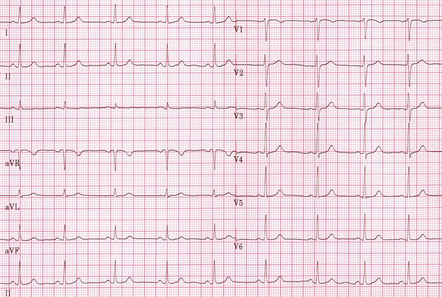


ECG strip with sinus rhythm

Sinus Rhythm

  • On the ECG, sinus rhythm is present
  • Notice how the height of the QRS complexes changes across different leads

What Does an ECG Diagnose?


Sources

  • ECG from Basics to Essentials Step by Step
  • litfl.com
  • ecgwaves.com
  • metealpaslan.com
  • medmastery.com
  • uptodate.com
  • ecgpedia.org
  • wikipedia.org
  • Strong Medicine
  • Understanding Pacemakers