ECGbook.com

Making Medical Education Free for All

ECGbook.com

Making Medical Education Free for All

Home /

Electrical Conduction System of the Heart

Electrical conduction system of the heart

Working Myocardium

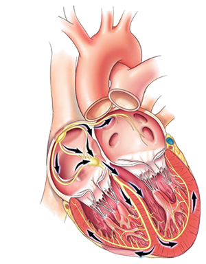
Heart electrical conduction system and cardiac muscle
  • The heart consists of 2 basic functional parts:
  • Working myocardium
    • It is the heart muscle
    • Contains myofibrils (muscle fibers)
    • Action potential of the myocardium has 5 phases
    • Impulse spreads through the working myocardium slowly
    • When the impulse reaches the myocardium, it triggers contraction
      • Results in atrial systole and ventricular systole

  • Heart conduction system
    • Consists of "electrical wires"
      • Along which the impulse travels from the SA node
    • Action potential of the conduction system has 3 phases
    • Impulse spreads through the conduction system quickly
      • Because it contains gap junctions
      • Through which ions quickly pass during impulse propagation
    • It does not have contractile ability (lacks myofibrils)

Electro-Mechanical Properties of the Heart

Heart myocardium and electrical conduction system - anatomy and physiology

Heart Conduction System

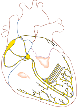
Parts of the heart electrical conduction system - anatomy and physiology
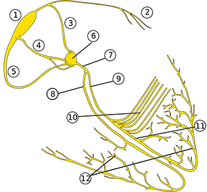
  • Supraventricular conduction system:
    • Sinoatrial (SA) Node (1)
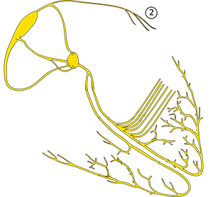
    • Bachmann's Bundle (2)
    • Internodal Pathways (3,4,5)
      • Anterior Internodal Pathway (James' Bundle) (3)
      • Middle Internodal Pathway (Wenckebach's Bundle) (4)
      • Posterior Internodal Pathway (Thorel's Bundle) (5)
    • Atrioventricular (AV) Junction (6,7)
      • Atrioventricular (AV) Node (6)
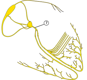
      • His Bundle (7)

  • Ventricular conduction system
    • Right Tawara's Branch (8)
    • Left Tawara's Branch (9)
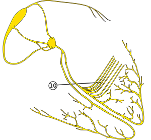
    • Left Posterior Fascicle (10)
    • Left Anterior Fascicle (11)
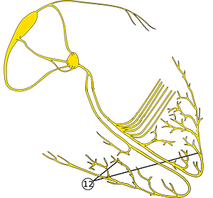
    • Purkinje Fibers (12)

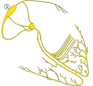
Sinoatrial (SA) Node (1)

Sinoatrial (SA) node and heart conduction electrical system

Bachmann's Bundle (2)

Bachmann's bundle and heart conduction electrical system
  • Emerges from the anterior internodal pathway
    • and continues to the left atrium
  • Transfers impulses from the SA node to the myocardium of the left atrium
  • Ensures parallel activation of the atria
  • If interrupted, it results in

Internodal Pathways (3, 4, 5)

Anterior (Wenckebach), middle (Thorel's), posterior internodal (James) tract and heart conduction electrical system

Atrioventricular (AV) Node (6)

Atrioventricular (AV) node and heart conduction electrical system
  • Atrioventricular (AV) Junction is the collective term for:
    • AV Node
    • His Bundle

  • The AV node is the only electrical connection between the atria and ventricles
  • The AV node is a secondary pacemaker
    • In case of SA node dysfunction
    • It begins to generate impulses with a frequency of 40-60/min.
    • Impulses from the AV node are seen on the ECG as an junctional rhythm
  • The AV node has 2 pathways:
    • Fast and slow
    • Physiologically, impulses pass only through the fast pathway
    • If impulses start to circulate (re-entry) through the pathways, AVNRT can occur
  • Impulse conduction through the AV node is decremental (slowed)
    • Conduction disturbances can lead to AV blocks

  • The AV node consists of 3 functional parts:
    • Transient Zone
      • Envelops the AV node (like a shell)
      • Composed of autonomic cells
      • It is the most arrhythmogenic part of the AV node
    • Compact Zone
      • Forms the core of the AV node
      • Histologically similar to the SA node
      • Slows down the impulse transmission to the ventricles and also acts as a filter
      • The secondary pacemaker is located in the compact zone
    • Trigger Zone
      • The impulse slows down in the compact zone
        • When it reaches the trigger zone, it speeds up
      • The impulse then quickly passes through the trigger zone to the His Bundle

His Bundle (7)

Bundle of His and heart conduction electrical system

Right Tawara Branch (8)

Right bundle branch and heart conduction electrical system
  • Begins in the area of the bifurcation of the His Bundle
  • Its terminal parts are Purkinje fibers
  • The impulse from the right branch depolarizes the entire right ventricle
  • A block can occur with right Tawara branch block (RBBB)

Left Tawara Branch (9)

Left bundle branch and heart conduction electrical system

Left Posterior Fascicle (10)

Left posterior fascicle and heart conduction electrical system
  • Begins at the bifurcation of the left Tawara branch
    • Ends in the posterior part of the left ventricle
  • Activates the posterior and upper parts of the left ventricle
  • The left posterior fascicle is not a single entity
    • It consists of a group of parallel fascicles
  • In case of disruption, a left posterior fascicular block occurs

Left Anterior Fascicle (11)

Left anterior fascicle and heart conduction electrical system

Purkinje Fibers (12)

Purkinje fibers and heart conduction electrical system
  • Form a dense network of fibers in the ventricles
  • Are the last parts of the conduction system in the ventricles, primarily originating from
    • The right bundle branch
    • The left posterior fascicle
    • The left anterior fascicle
  • Fibers transmit impulses to the subendocardium
  • Purkinje fibers act as a tertiary pacemaker
    • Activate in the event of dysfunction in the SA and AV nodes
    • Generate impulses with a frequency of 20-40/min.
    • On the ECG, this generates a ventricular rhythm


Sources

  • ECG from Basics to Essentials Step by Step
  • litfl.com
  • ecgwaves.com
  • metealpaslan.com
  • medmastery.com
  • uptodate.com
  • ecgpedia.org
  • wikipedia.org
  • Strong Medicine
  • Understanding Pacemakers


šípka späť

Electrical Conduction System of the Heart

Electrical Conduction System of the Heart

Working Myocardium

  • The heart consists of 2 basic functional parts:
  • Working myocardium
    • It is the heart muscle
    • Contains myofibrils (muscle fibers)
    • Action potential of the myocardium has 5 phases
    • Impulse spreads through the working myocardium slowly
    • When the impulse reaches the myocardium, it triggers contraction
      • Results in atrial systole and ventricular systole

  • Heart conduction system
    • Consists of "electrical wires"
      • Along which the impulse travels from the SA node
    • Action potential of the conduction system has 3 phases
    • Impulse spreads through the conduction system quickly
      • Because it contains gap junctions
      • Through which ions quickly pass during impulse propagation
    • It does not have contractile ability (lacks myofibrils)


Heart electrical conduction system and cardiac muscle

Electro-Mechanical Properties of the Heart

  • Automaticity
    • Heart cells spontaneously generate action potentials (impulses)
    • Physiologically, impulses with the highest frequency are generated in the SA node
      • The heart can beat for a while even outside the body
  • Excitability
    • It is the ability of heart cells to respond to impulses
    • An impulse from the SA node activates adjacent cells
  • Conductivity
    • The impulse quickly spreads from cell to cell through the conduction system
      • and then more slowly through the working myocardium
    • The impulse thus spreads from the SA node
      • through the entire conduction system and then to the myocardium
  • Contractility
    • It is the ability of contraction
    • This property applies only to working myocardium
      • Cells of the conduction system do not contract
    • The working myocardium contains myofibrils (muscle fibers)
    • When an impulse reaches the working myocardium
      • it results in atrial systole and ventricular systole
Heart myocardium and electrical conduction system - anatomy and physiology

Heart Conduction System

  • Supraventricular conduction system:
    • Sinoatrial (SA) Node (1)
    • Bachmann's Bundle (2)
    • Internodal Pathways (3,4,5)
      • Anterior Internodal Pathway (James' Bundle) (3)
      • Middle Internodal Pathway (Wenckebach's Bundle) (4)
      • Posterior Internodal Pathway (Thorel's Bundle) (5)
    • Atrioventricular (AV) Junction (6,7)
      • Atrioventricular (AV) Node (6)
      • His Bundle (7)

  • Ventricular conduction system
    • Right Tawara's Branch (8)
    • Left Tawara's Branch (9)
    • Left Posterior Fascicle (10)
    • Left Anterior Fascicle (11)
    • Purkinje Fibers (12)


Parts of the heart electrical conduction system - anatomy and physiology

Sinoatrial (SA) Node (1)

  • Located in the right atrium (measuring 15x3mm)
  • Spontaneously generates impulses with a frequency of 60-100/min.
  • It is the primary pacemaker
    • Because it generates impulses with the highest frequency
    • All other parts generate impulses at a lower frequency
    • The SA node deactivates ectopic foci

Sinoatrial (SA) node and heart conduction electrical system

Bachmann's Bundle (2)

  • Emerges from the anterior internodal pathway
    • and continues to the left atrium
  • Transfers impulses from the SA node to the myocardium of the left atrium
  • Ensures parallel activation of the atria
  • If interrupted, it results in

Bachmann's bundle and heart conduction electrical system

Internodal Pathways (3, 4, 5)

  • SA node and AV node are connected by 3 internodal pathways:
    • Anterolateral Internodal Pathway (James' Bundle) (3)
    • Middle Internodal Pathway (Wenckebach's Bundle) (4)
    • Posterior Internodal Pathway (Thorel's Bundle) (5)
  • Typically, only 1 pathway is connected to the AV node
    • The other 2 pathways end before the AV node in the atrium
    • Only 10% of the population has 2 pathways connected to the AV node
  • If 2 pathways enter the AV node, then
    • one is faster and the other is slower
    • The AV node is activated always via the faster pathway
      • The impulse from the faster pathway resets the impulse from the slower pathway
      • (refractory period of the action potential)

Anterior (Wenckebach), middle (Thorel's), posterior internodal (James) tract and heart conduction electrical system

Atrioventricular (AV) Node (6)

  • Atrioventricular (AV) Junction is the collective term for:
    • AV Node
    • His Bundle

  • The AV node is the only electrical connection between the atria and ventricles
  • The AV node is a secondary pacemaker
    • In case of SA node dysfunction
    • It begins to generate impulses with a frequency of 40-60/min.
    • Impulses from the AV node are seen on the ECG as an junctional rhythm
  • The AV node has 2 pathways:
    • Fast and slow
    • Physiologically, impulses pass only through the fast pathway
    • If impulses start to circulate (re-entry) through the pathways, AVNRT can occur
  • Impulse conduction through the AV node is decremental (slowed)
    • Conduction disturbances can lead to AV blocks

  • The AV node consists of 3 functional parts:
    • Transient Zone
      • Envelops the AV node (like a shell)
      • Composed of autonomic cells
      • It is the most arrhythmogenic part of the AV node
    • Compact Zone
      • Forms the core of the AV node
      • Histologically similar to the SA node
      • Slows down the impulse transmission to the ventricles and also acts as a filter
      • The secondary pacemaker is located in the compact zone
    • Trigger Zone
      • The impulse slows down in the compact zone
        • When it reaches the trigger zone, it speeds up
      • The impulse then quickly passes through the trigger zone to the His Bundle

Atrioventricular (AV) node and heart conduction electrical system

His Bundle (7)

  • Starts in the terminal part of the right atrium
  • Passes through the atrioventricular septum
  • Ends in the interventricular septum

  • The His Bundle transmits the impulse from the AV node to the Tawara branches
  • The impulse emerges from the His Bundle to the ventricular septum
    • On the ECG, this produces a Q wave

Bundle of His and heart conduction electrical system

Right Tawara Branch (8)

  • Begins in the area of the bifurcation of the His Bundle
  • Its terminal parts are Purkinje fibers
  • The impulse from the right branch depolarizes the entire right ventricle
  • A block can occur with right Tawara branch block (RBBB)

Right bundle branch and heart conduction electrical system

Left Tawara Branch (9)

  • Begins in the area of the bifurcation of the His Bundle
  • Its terminal part divides into
    • Left anterior fascicle
    • Left posterior fascicle
  • Purkinje fibers extend from the fascicles
  • A block can occur with left Tawara branch block (LBBB)

Left bundle branch and heart conduction electrical system

Left Posterior Fascicle (10)

  • Begins at the bifurcation of the left Tawara branch
    • Ends in the posterior part of the left ventricle
  • Activates the posterior and upper parts of the left ventricle
  • The left posterior fascicle is not a single entity
    • It consists of a group of parallel fascicles
  • In case of disruption, a left posterior fascicular block occurs

Left posterior fascicle and heart conduction electrical system

Left Anterior Fascicle (11)

  • Begins at the bifurcation of the left Tawara branch
    • Ends in the anterior part of the left ventricle
  • Activates the lower and anterior parts of the left ventricle
  • The left anterior fascicle is a single entity
  • Left anterior fascicular block
    • Is more common than the posterior fascicular block
    • Because it only requires one anterior fascicle to be disrupted for a block to occur

Left anterior fascicle and heart conduction electrical system

Purkinje Fibers (12)

  • Form a dense network of fibers in the ventricles
  • Are the last parts of the conduction system in the ventricles, primarily originating from
    • The right bundle branch
    • The left posterior fascicle
    • The left anterior fascicle
  • Fibers transmit impulses to the subendocardium
  • Purkinje fibers act as a tertiary pacemaker
    • Activate in the event of dysfunction in the SA and AV nodes
    • Generate impulses with a frequency of 20-40/min.
    • On the ECG, this generates a ventricular rhythm

Purkinje fibers and heart conduction electrical system


Sources

  • ECG from Basics to Essentials Step by Step
  • litfl.com
  • ecgwaves.com
  • metealpaslan.com
  • medmastery.com
  • uptodate.com
  • ecgpedia.org
  • wikipedia.org
  • Strong Medicine
  • Understanding Pacemakers