ECGbook.com

Making Medical Education Free for All

ECGbook.com

Making Medical Education Free for All

Home /

Hyperventilation-induced T Wave Inversion

Hyperventilation induced T wave inversion

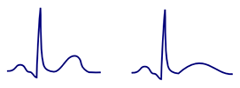
Normal T Wave

ECG normal positive T wave

Concordant T Wave


ECG abnormal disconcordance T wave, positive QRS, negative inverted T wave

Disconcordant T Wave


  • The T wave is the most variable wave on the ECG
  • It is concordant (has the same polarity as the QRS)
    • Positive QRS - positive T wave
    • Negative QRS - negative T wave
  • It is positive (I, II, V3-V6)
    • It may be isolated negative (III, aVL)
  • It is negative (aVR)
  • Amplitude
  • For ECG diagnosis, the dynamics of the T wave is important
    • If the T wave remains the same - it often does not indicate a serious condition
    • If the T wave changes - it often indicates a serious condition (ischemia)

Hyperventilation

Hyperventilation, overbreathing, Hyperventilation syndrome, respiratory alkalosis

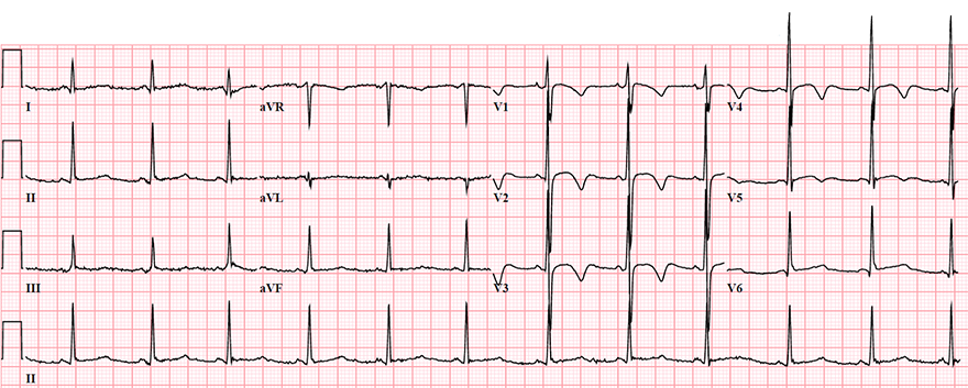
Inverted T Waves and Hyperventilation

Hyperventilation T wave inversion
  • Hyperventilation causes (temporary changes) on an EKG:
  • The mechanism of EKG changes is primarily transient hypokalemia
    • Which results from hyperventilation-induced respiratory alkalosis (Blood pH increases)
    • EKG changes persist for approximately 1 minute after hyperventilation

Differential Diagnosis


ECG T wave inversion, hyperventilation

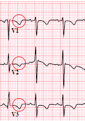
Persistent Juvenile T Waves

  • It involved a 24-year-old woman who was frightened and hyperventilating, and she had no structural heart disease
  • Inverted T waves (V1-V5)
  • After calming the patient, these T waves resolved


ECG persistent juvenile T wave pattern, african-american female

Persistent Juvenile T Waves


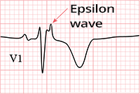
ECG epsilon wave, inverted T wave

ECG (V1-V3) arrhythmogenic right ventricular hypertrophy, epsilon wave, inverted T wave

Arrhythmogenic Right Ventricular Dysplasia
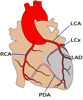


Heart stenosis: Circumflex branch of left coronary artery (LCx), Anterior interventricular branch of left coronary artery (LAD)
ECG ischemic inverted T waves, unstable angina pectoris, stenosis LAD, LCx

Inverted Ischemic T Waves and Unstable Angina Pectoris


Sources

  • ECG from Basics to Essentials Step by Step
  • litfl.com
  • ecgwaves.com
  • metealpaslan.com
  • medmastery.com
  • uptodate.com
  • ecgpedia.org
  • wikipedia.org
  • Strong Medicine
  • Understanding Pacemakers


šípka späť

Hyperventilation-induced T Wave Inversion

Hyperventilation induced T wave inversion

Normal T Wave

  • The T wave is the most variable wave on the ECG
  • It is concordant (has the same polarity as the QRS)
    • Positive QRS - positive T wave
    • Negative QRS - negative T wave
  • It is positive (I, II, V3-V6)
    • It may be isolated negative (III, aVL)
  • It is negative (aVR)
  • Amplitude
  • For ECG diagnosis, the dynamics of the T wave is important
    • If the T wave remains the same - it often does not indicate a serious condition
    • If the T wave changes - it often indicates a serious condition (ischemia)
ECG normal positive T wave

Concordant T Wave


ECG abnormal disconcordance T wave, positive QRS, negative inverted T wave

Disconcordant T Wave


Hyperventilation

  • During hyperventilation, more carbon dioxide is exhaled
    • than is produced in the body
  • Respiratory alkalosis occurs (pH increases in the blood)
    • There is a change in mineral concentration
  • Ionized calcium decreases
    • Muscular excitability increases and tetanic contractions occur
      • Obstetric hand sign – Trousseau's sign
      • Carpopedal spasms – Chvostek's sign
  • An increase in pH by 0.1 causes a decrease in potassium by approximately 0.5mmol/l
  • If serious conditions are excluded (e.g., pulmonary embolism)
    • then treatment of hyperventilation includes:
      • Calming the patient
      • Breathing into a plastic bag (the patient rebreathes carbon dioxide)


Hyperventilation, overbreathing, Hyperventilation syndrome, respiratory alkalosis

Inverted T Waves and Hyperventilation

  • Hyperventilation causes (temporary changes) on an EKG:
  • The mechanism of EKG changes is primarily transient hypokalemia
    • Which results from hyperventilation-induced respiratory alkalosis (Blood pH increases)
    • EKG changes persist for approximately 1 minute after hyperventilation

Hyperventilation T wave inversion

Differential Diagnosis


ECG T wave inversion, hyperventilation

Persistent Juvenile T Waves

  • It involved a 24-year-old woman who was frightened and hyperventilating, and she had no structural heart disease
  • Inverted T waves (V1-V5)
  • After calming the patient, these T waves resolved


ECG persistent juvenile T wave pattern, african-american female

Persistent Juvenile T Waves


ECG (V1-V3) arrhythmogenic right ventricular hypertrophy, epsilon wave, inverted T wave

Arrhythmogenic Right Ventricular Dysplasia

ECG epsilon wave, inverted T wave


ECG ischemic inverted T waves, unstable angina pectoris, stenosis LAD, LCx

Inverted Ischemic T Waves and Unstable Angina Pectoris

Heart stenosis: Circumflex branch of left coronary artery (LCx), Anterior interventricular branch of left coronary artery (LAD)


Sources

  • ECG from Basics to Essentials Step by Step
  • litfl.com
  • ecgwaves.com
  • metealpaslan.com
  • medmastery.com
  • uptodate.com
  • ecgpedia.org
  • wikipedia.org
  • Strong Medicine
  • Understanding Pacemakers