ECGbook.com

Making Medical Education Free for All

ECGbook.com

Making Medical Education Free for All

Home /

Idiopathic Ventricular Tachycardia

Idiopathic ventricular tachycardia

Ventricular Tachycardia

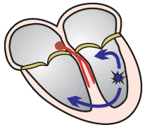
Ectopic focus, exit site, broad QRS complex, ventricular tachycardia

Idiopathic Ventricular Tachycardia

idiopathic monomorphic ventricular tachycardia, RVOT-VT, LVOT-VT, Fascicular VT

Ventricular Tachycardias from Outflow Tracts


Ventricular Tachycardia from the Right Ventricular Outflow Tract
Right Ventricular Outflow Tract (RVOT) Tachycardia (Monomorphic VT)

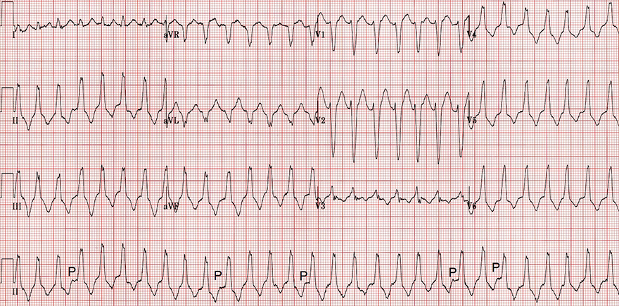
ECG Right ventricular outflow tract (RVOT) ventricular tachycardia, Inferior axis, AV dissociation

Ventricular Tachycardia from the Right Ventricular Outflow Tract

  • Frequency: 160/min.
  • Wide QRS complexes (>0.12s)
  • Transition Zone (V3)
    • The position of the transition zone differentiates VT from the right and left outflow tracts
  • Pattern of left bundle branch block
    • Wide QRS complexes
    • Deep S in V1
    • Dominant R in V6
  • Vertical Axis (+ 90°)
    • Positive QRS in II, III, aVF
  • AV Dissociation
    • P waves visible in continuous lead II, deforming the QRS complexes
    • AV dissociation is a fundamental sign of ventricular tachycardia

Ventricular Tachycardia from the Left Ventricular Outflow Tract
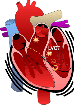
Idiopathic Left Ventricular Outflow Tract (LVOT) Tachycardia

ECG Idiopathic  Left Ventricular Outflow Tract (LVOT) Tachycardia

Ventricular Tachycardia from the Left Ventricular Outflow Tract

  • Heart Rate: 140/min.
  • Wide QRS complexes (>0.12s)
  • Pattern of left bundle branch block
    • Wide QRS complexes
    • Deep S wave (V1)
    • Dominant R wave (V6)
  • Vertical axis (+100°)
    • Positive QRS in (II, III, aVF)
  • Transition zone (V3)
    • It is, however, VT from the left ventricular outflow tract due to the presence of additional features:
      • Wide R wave in V1 (more than 50% of QRS width)
      • Higher R wave in V1 (more than 30% of S wave)

Fascicular Ventricular Tachycardia

Posterior fascicular VT, Anterior fascicular VT, Upper septal fascicular VT

ECG Anterior fascicular VT, RBBB morphology, Right axis deviation,

Anterior Fascicular Ventricular Tachycardia

  • Heart rate: 150/min.
  • QRS complexes are borderline wide (0.12s)
  • Right Bundle Branch Block (RBBB)
    • rSR' in V1 - The right rabbit ear is larger
    • Deep and wide S wave in V6
  • Left Posterior Hemiblock (axis deviation right +120º)
    • Negative QRS in I
    • Positive QRS in aVF, III
  • May be a supraventricular tachycardia with bifascicular block (RBBB + left posterior hemiblock)
    • However, the tachycardia continued after adenosine administration
    • Adenosine slows down the AV node and terminates supraventricular tachycardia
  • The tachycardia resolved after administration of verapamil
    • This is a verapamil-sensitive tachycardia, which is characteristic of Fascicular VT
  • This is Fascicular VT (re-entry is in the left anterior fascicle)
    • Even though typical signs of ventricular tachycardia are absent


ECG Posterior fascicular VT, RBBB morphology, left axis deviation, AV dissociation, fusion beat, capture beat

Posterior Fascicular Ventricular Tachycardia


Sources

  • ECG from Basics to Essentials Step by Step
  • litfl.com
  • ecgwaves.com
  • metealpaslan.com
  • medmastery.com
  • uptodate.com
  • ecgpedia.org
  • wikipedia.org
  • Strong Medicine
  • Understanding Pacemakers


šípka späť

Idiopathic Ventricular Tachycardia

Idiopathic ventricular tachycardia

Ventricular Tachycardia


Ectopic focus, exit site, broad QRS complex, ventricular tachycardia

Idiopathic Ventricular Tachycardia


idiopathic monomorphic ventricular tachycardia, RVOT-VT, LVOT-VT, Fascicular VT

Ventricular Tachycardias from Outflow Tracts


Ventricular Tachycardia from the Right Ventricular Outflow Tract


Right Ventricular Outflow Tract (RVOT) Tachycardia (Monomorphic VT)


ECG Right ventricular outflow tract (RVOT) ventricular tachycardia, Inferior axis, AV dissociation

Ventricular Tachycardia from the Right Ventricular Outflow Tract

  • Frequency: 160/min.
  • Wide QRS complexes (>0.12s)
  • Transition Zone (V3)
    • The position of the transition zone differentiates VT from the right and left outflow tracts
  • Pattern of left bundle branch block
    • Wide QRS complexes
    • Deep S in V1
    • Dominant R in V6
  • Vertical Axis (+ 90°)
    • Positive QRS in II, III, aVF
  • AV Dissociation
    • P waves visible in continuous lead II, deforming the QRS complexes
    • AV dissociation is a fundamental sign of ventricular tachycardia

Ventricular Tachycardia from the Left Ventricular Outflow Tract

  • Left Ventricular Outflow Tract (LVOT) Tachycardia

  • Ventricular Tachycardia from the Left Ventricular Outflow Tract
  • Accounts for 20% of idiopathic VTs
  • Common sites of origin:
    • Aortic root near the origin of the coronary arteries (Valsalva sinus)
    • Basal part of the left ventricle
    • Epicardium of the left ventricular outflow tract
  • Main vector points downwards towards the left ventricle
    • Does not point towards the right ventricle because the left ventricle is more massive

  • On the ECG, it resembles the pattern of VT from the right ventricular outflow tract:
    • Pattern of left bundle branch block
    • Positive QRS in inferior leads (II, III, aVF)
    • VT from the left ventricular outflow tract has additional features:
      • Wide R wave in V1-2 (more than 50% of QRS width)
      • Higher R wave in V1-2 (more than 30% of S wave)
      • Transition zone ≤ V2
      • Notch in the ascending part of the R wave (V4-6)
Idiopathic Left Ventricular Outflow Tract (LVOT) Tachycardia


ECG Idiopathic  Left Ventricular Outflow Tract (LVOT) Tachycardia

Ventricular Tachycardia from the Left Ventricular Outflow Tract

  • Heart Rate: 140/min.
  • Wide QRS complexes (>0.12s)
  • Pattern of left bundle branch block
    • Wide QRS complexes
    • Deep S wave (V1)
    • Dominant R wave (V6)
  • Vertical axis (+100°)
    • Positive QRS in (II, III, aVF)
  • Transition zone (V3)
    • It is, however, VT from the left ventricular outflow tract due to the presence of additional features:
      • Wide R wave in V1 (more than 50% of QRS width)
      • Higher R wave in V1 (more than 30% of S wave)

Fascicular Ventricular Tachycardia

  • Fascicular VT is very rare
    • It constitutes 10% of idiopathic VTs
  • Mechanism is re-entry
    • Re-entry passes through one fascicle and the adjacent myocardium
      • Fast pathway is formed by the fascicle (anterior or posterior)
      • Slow pathway is formed by the adjacent myocardium near the fascicle
  • Occurs mainly in men aged 15-40 years
  • Referred to as verapamil-sensitive ventricular tachycardia
    • Responds well (terminates) after administration of verapamil

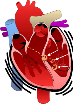
  • According to the location of re-entry, there are 3 types of fascicular VTs:
  • Beware of interfascicular VT
    • Occurs in structurally damaged hearts (not idiopathic)
    • Re-entry passes through both the anterior and posterior fascicles
      • The fast pathway is in one fascicle
      • The slow pathway is in the other fascicle

Posterior fascicular VT, Anterior fascicular VT, Upper septal fascicular VT


ECG Anterior fascicular VT, RBBB morphology, Right axis deviation,

Anterior Fascicular Ventricular Tachycardia

  • Heart rate: 150/min.
  • QRS complexes are borderline wide (0.12s)
  • Right Bundle Branch Block (RBBB)
    • rSR' in V1 - The right rabbit ear is larger
    • Deep and wide S wave in V6
  • Left Posterior Hemiblock (axis deviation right +120º)
    • Negative QRS in I
    • Positive QRS in aVF, III
  • May be a supraventricular tachycardia with bifascicular block (RBBB + left posterior hemiblock)
    • However, the tachycardia continued after adenosine administration
    • Adenosine slows down the AV node and terminates supraventricular tachycardia
  • The tachycardia resolved after administration of verapamil
    • This is a verapamil-sensitive tachycardia, which is characteristic of Fascicular VT
  • This is Fascicular VT (re-entry is in the left anterior fascicle)
    • Even though typical signs of ventricular tachycardia are absent


ECG Posterior fascicular VT, RBBB morphology, left axis deviation, AV dissociation, fusion beat, capture beat

Posterior Fascicular Ventricular Tachycardia


Sources

  • ECG from Basics to Essentials Step by Step
  • litfl.com
  • ecgwaves.com
  • metealpaslan.com
  • medmastery.com
  • uptodate.com
  • ecgpedia.org
  • wikipedia.org
  • Strong Medicine
  • Understanding Pacemakers