|
|
ECGbook.com Making Medical Education Free for All |
|
|
ECGbook.com Making Medical Education Free for All |
|
|
ECGbook.com Making Medical Education Free for All |

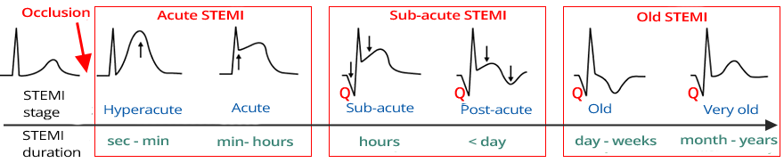
STEMI Stages by Phase


Subacute Inferior STEMI

Old Inferior STEMI
Acute Inferior STEMI and Right Ventricular STEMI
Acute Inferior and Posterior STEMI
Acute Inferior and Posterior STEMI

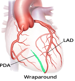
Acute Anterior and Inferior STEMI (Wraparound)

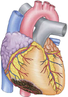
Coronary Angiography
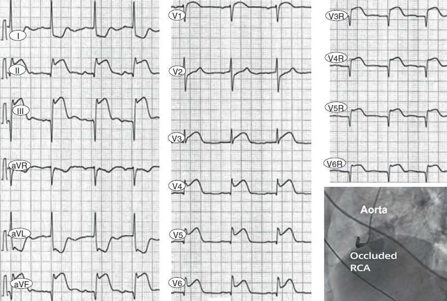
Acute Inferior STEMI and Right Ventricular STEMI
Sources
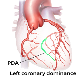
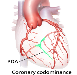
Coronary Artery Dominance
|
|
|
|
|
|
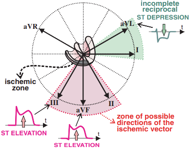
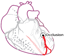
Inferior Wall Myocardial Infarction
|
|
ECG and Inferior Wall STEMI
|
|
STEMI Stages by Phase
Occlusion of the Right Coronary Artery
|
|
ECG and Occlusion of the Dominant RCA
|
|
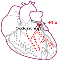
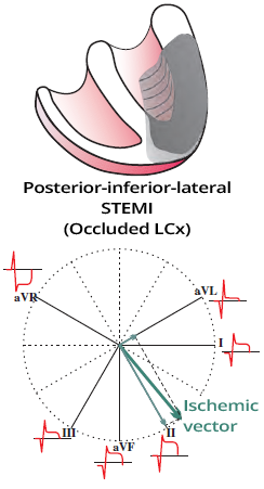
Occlusion of the Left Coronary Artery (LCx)
|
|
ECG and Occlusion of the Dominant LCx (Left Circumflex Artery)
|
|
Occlusion of the RIA in "Wraparound"
|
|
|
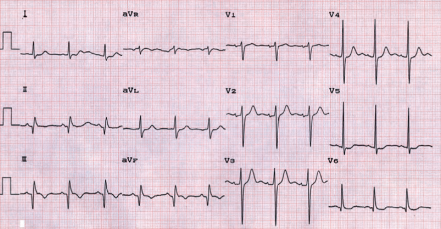
Subacute Inferior STEMI
|
|
|
Old Inferior STEMI
|
|
|
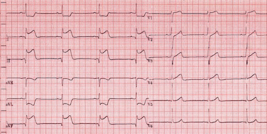
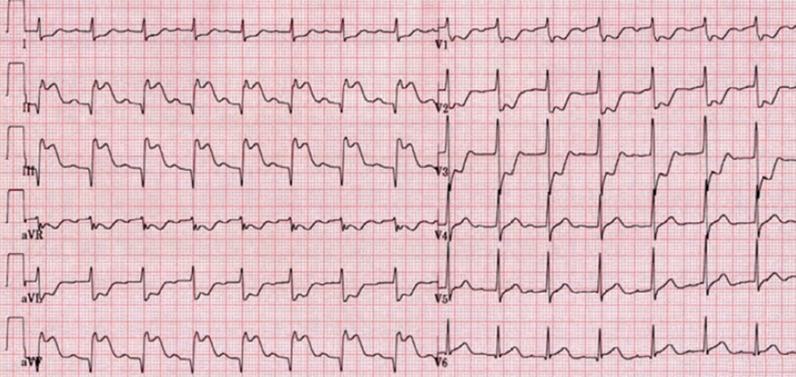
Acute Inferior STEMI and Right Ventricular STEMI
|
|
|
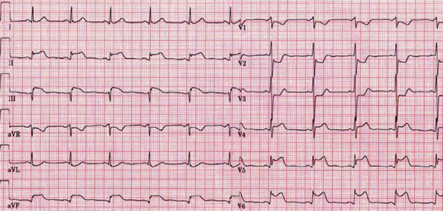
Acute Inferior and Posterior STEMI
|
|
|
Acute Inferior and Posterior STEMI
|
|
|
Acute Anterior and Inferior STEMI (Wraparound)
|
|
|
|
Coronary Angiography
|
|
Acute Inferior STEMI and Right Ventricular STEMI
|
|
Sources