ECGbook.com

Making Medical Education Free for All

ECGbook.com

Making Medical Education Free for All

Home /

J Wave (Osborn Wave)

J wave, Osborn wave

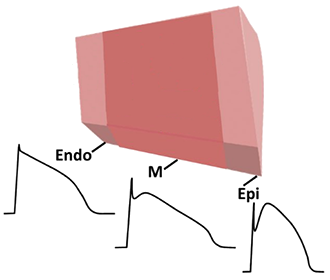
Action Potential and Myocardial Wall

Action potential in endocardium, epicardium, and myocardium
  • Action potential (AP) in the myocardial wall
    • Varies slightly in
      • Epicardium
      • Mid-myocardium (M-cells)
      • Endocardium


  • AP also varies in
    • right and left ventricles

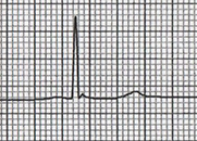
Normal ECG Curve

Normal QRS complex

J Wave Mechanism

ECG J wave mechanism action potential endocardium, epicardium
  • Changes in ion channels
    • During the Phase 1 of the action potential
    • Result in a change in the action potential curve
    • Between the endocardium and epicardium

  • Altered action potential curves
    • Generate a J wave on the ECG

Causes of J Wave (Osborn Wave)


ECG and J Wave (Osborn Wave)

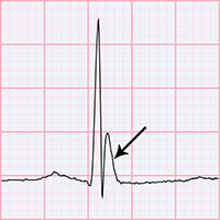
  • It is a narrow, peaked wave after the QRS complex
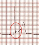
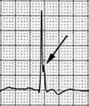
    • "Notch" on the terminal part of the QRS is a less common variant of the Osborn wave
  • Sometimes it may resemble ST elevations, but the Osborn wave is narrow and peaked
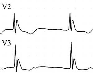
  • It is primarily seen in precordial leads (V1-V6)
    • It must be present in at least two adjacent leads
  • Not found in aVR or V1
ECG J wave, Osborn wave, camel-hump sign, late delta wave, hathook junction, hypothermic wave, K wave, H wave or current of injury


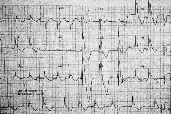
ECG J (Osborn) wave and hypothermia

Hypothermia


ECG benign early repolarization and J, Osborn wave

Benign Early Repolarization


ECG hypercalcemia and J wave as notch

Hypercalcemia


Hypothermia and the Osborn Wave

  • The most common cause of the Osborn wave is hypothermia < 32°C
  • Amplitude and width increase with worsening hypothermia
  • After correcting hypothermia, the Osborn wave may persist for 12-24 hours.
  • Any Osborn wave is a risk factor for ventricular fibrillation (Le syndrome d’Haïssaguerre)
    • Fibrillation most commonly occurs during rewarming of the body at 28-32°C

Hypothermia, ECG J wave, Osborn wave duration and amplitude

Osborn Wave and Hypothermia

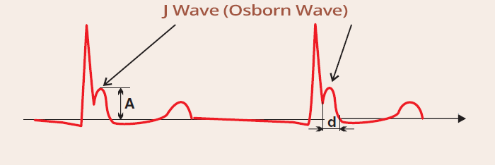
  • The amplitude (A) and width (d) of the Osborn wave increase with deepening hypothermia

ECG and Dynamics of the Osborn Wave in Hypothermia

ECG J wave (Osborn) and hypothermia 32 degrees Celsius

Hypothermia (32°C)


ECG J wave (Osborn) and hypothermia 34 degrees Celsius

Hypothermia (34°C)


ECG J wave (Osborn) and hypothermia 35 degrees Celsius

Hypothermia (35°C)


ECG J wave (Osborn) and hypothermia 36 degrees Celsius

Hypothermia (36°C)


Dynamics of the Osborn Wave in Hypothermia

ECG hypothermia and J wave, Osborn wave 27 degrees Celsius

Hypothermia (27°C)

  • Large Osborn waves (V2-6, I, II, III, aVF)

ECG hypothermia and J wave, Osborn wave 30 degrees Celsius

Hypothermia (30°C)

  • Temperature increase by 3°C
  • Notice how the Osborn wave has decreased

ECG hypothermic wave, J wave, Osborn wave, hypothermia 32 degrees Celsius

Hypothermia (32.5°C)

  • Temperature increase by 2.5°C
  • Small Osborn wave just after the QRS complex


ECG hypothermic J wave, hypothermia 29 degree

J Wave and Hypothermia (29°C)


ECG J wave - notch, and hypercalcaemia

J Wave and Hypercalcemia

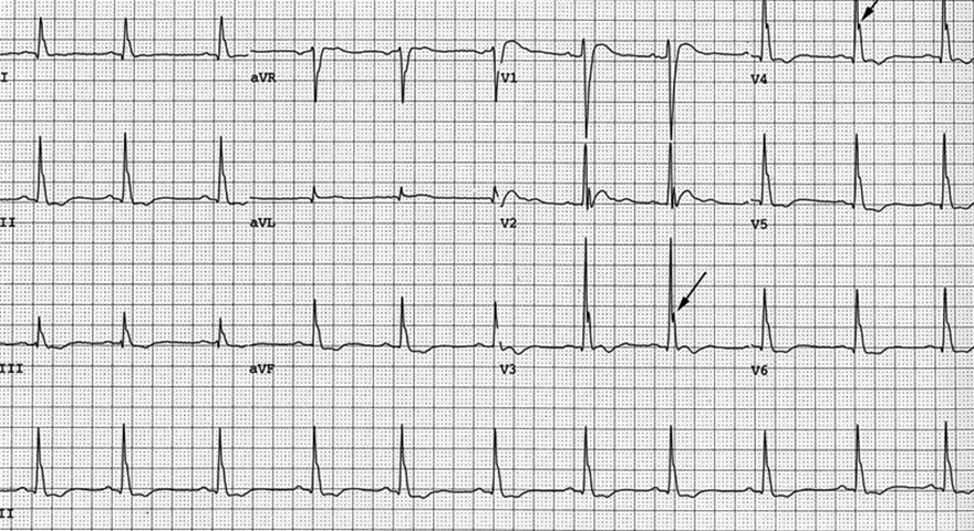
  • Osborn waves (arrows)
  • Osborn waves create a notch at the terminal part of the QRS complex (less common variant of the Osborn wave)
  • The patient had severe hypercalcemia of 4.1 mmol/l


ECG benign early repolarisation, hathook junction, J wave, late delta wave

J Wave and Benign Early Repolarization


Sources

  • ECG from Basics to Essentials Step by Step
  • litfl.com
  • ecgwaves.com
  • metealpaslan.com
  • medmastery.com
  • uptodate.com
  • ecgpedia.org
  • wikipedia.org
  • Strong Medicine
  • Understanding Pacemakers


šípka späť

J Wave (Osborn Wave)

J wave, Osborn wave

Action Potential and Myocardial Wall

Action potential in endocardium, epicardium, and myocardium
  • Action potential (AP) in the myocardial wall
    • Varies slightly in
      • Epicardium
      • Mid-myocardium (M-cells)
      • Endocardium


  • AP also varies in
    • right and left ventricles

Normal ECG Curve

Normal QRS complex

  • Action Potential
    • Differs slightly between the endocardium and epicardium
    • Due to differences in ion channel properties
  • Phase 0 and 1 generate the QRS complex on the ECG

J Wave Mechanism

ECG J wave mechanism action potential endocardium, epicardium
  • Changes in ion channels
    • During the Phase 1 of the action potential
    • Result in a change in the action potential curve
    • Between the endocardium and epicardium

  • Altered action potential curves
    • Generate a J wave on the ECG

Causes of J Wave (Osborn Wave)


ECG and J Wave (Osborn Wave)

  • It is a narrow, peaked wave after the QRS complex
    • "Notch" on the terminal part of the QRS is a less common variant of the Osborn wave
  • Sometimes it may resemble ST elevations, but the Osborn wave is narrow and peaked
  • It is primarily seen in precordial leads (V1-V6)
    • It must be present in at least two adjacent leads
  • Not found in aVR or V1

ECG J wave, Osborn wave, camel-hump sign, late delta wave, hathook junction, hypothermic wave, K wave, H wave or current of injury


ECG J (Osborn) wave and hypothermia ECG benign early repolarization and J, Osborn wave ECG hypercalcemia and J wave as notch

Hypothermia

  • Osborn wave at the end of QRS
  • in two adjacent leads

Benign Early Repolarization

Hypercalcemia
  • Osborn wave as a
    notch (rare variant)

Hypothermia and the Osborn Wave

  • The most common cause of the Osborn wave is hypothermia < 32°C
  • Amplitude and width increase with worsening hypothermia
  • After correcting hypothermia, the Osborn wave may persist for 12-24 hours.
  • Any Osborn wave is a risk factor for ventricular fibrillation (Le syndrome d’Haïssaguerre)
    • Fibrillation most commonly occurs during rewarming of the body at 28-32°C

Hypothermia, ECG J wave, Osborn wave duration and amplitude

Osborn Wave and Hypothermia

  • The amplitude (A) and width (d) of the Osborn wave increase with deepening hypothermia

ECG and Dynamics of the Osborn Wave in Hypothermia

ECG J wave (Osborn) and hypothermia 32 degrees Celsius

Hypothermia (32°C)


ECG J wave (Osborn) and hypothermia 34 degrees Celsius

Hypothermia (34°C)


ECG J wave (Osborn) and hypothermia 35 degrees Celsius

Hypothermia (35°C)


ECG J wave (Osborn) and hypothermia 36 degrees Celsius

Hypothermia (36°C)


Dynamics of the Osborn Wave in Hypothermia

ECG hypothermia and J wave, Osborn wave 27 degrees Celsius

Hypothermia (27°C)

  • Large Osborn waves (V2-6, I, II, III, aVF)

ECG hypothermia and J wave, Osborn wave 30 degrees Celsius

Hypothermia (30°C)

  • Temperature increase by 3°C
  • Notice how the Osborn wave has decreased

ECG hypothermic wave, J wave, Osborn wave, hypothermia 32 degrees Celsius

Hypothermia (32.5°C)

  • Temperature increase by 2.5°C
  • Small Osborn wave just after the QRS complex


ECG hypothermic J wave, hypothermia 29 degree

J Wave and Hypothermia (29°C)


ECG J wave - notch, and hypercalcaemia

J Wave and Hypercalcemia

  • Osborn waves (arrows)
  • Osborn waves create a notch at the terminal part of the QRS complex (less common variant of the Osborn wave)
  • The patient had severe hypercalcemia of 4.1 mmol/l


ECG benign early repolarisation, hathook junction, J wave, late delta wave

J Wave and Benign Early Repolarization


Sources
  • ECG from Basics to Essentials Step by Step
  • litfl.com
  • ecgwaves.com
  • metealpaslan.com
  • medmastery.com
  • uptodate.com
  • ecgpedia.org
  • wikipedia.org
  • Strong Medicine
  • Understanding Pacemakers