ECGbook.com

Making Medical Education Free for All

ECGbook.com

Making Medical Education Free for All

Home /

Junctional Rhythm

AV junctional rhythm, Junctional rhythm, Junctional escape rhythm

Heart Rhythm

heart rhythm mechanism, sinus rhythm, junctional rhythm, ventricular rhythm

Basic Heart Rhythms

  • Heart rhythm is determined by the site (sites) that generates impulses with the highest frequency (overdrive suppression)
  • There are 3 basic heart rhythms
  • Sinus rhythm is the physiological heart rhythm
  • Junctional rhythm is a secondary pacemaker
    • The AV junction generates impulses with the second highest frequency after the SA node
    • Junctional rhythm occurs if
      • impulses from the atria cease to arrive at the AV junction
      • the AV junction starts generating impulses with a higher frequency than the atria

Junctional Rhythm


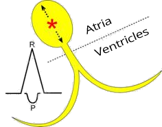
AV node, AV junction depolarization

Junctional Rhythm - Most Common Causes

Sinus Rhythm and Junctional Rhythm

SA node and sinus rhythm
ECG sinus rhythm vs. junctional rhythm

Sinus Rhythm


AV node, AV junction
ECG junctional rhythm vs. sinus rhythm

Junctional Rhythm

  • Impulses are generated by the AV junction with a frequency of: 40-60/min.
  • From the AV junction, the impulse spreads:
    • to both the atria and the ventricles
  • Ventricular vector moves toward lead II
    • A QRS complex is formed
  • Atrial vector moves toward the SA node
    • Direction is away from lead II
    • A negative - retrograde P wave is formed
  • The ventricles are activated first (QRS) and then the atria (negative P wave)

P Wave and Junctional Rhythm

atria upwards (P waves) and ventricles downwards (QRS complex)
upper AV nodal junctional rhythm and retrograde P wave
Upper Junctional Rhythm
middle AV nodal junctional rhythm
Middle Junctional Rhythm
down AV nodal junctional rhythm and retrograde P wave
Lower Junctional Rhythm

Upper Junctional Rhythm

upper AV junctional rhythm, retrograde P wave before QRS complex
  • Impulses originate in the upper part of the AV junction
    • Each impulse simultaneously spreads to both the atria and the ventricles
    • Atria are activated before the ventricles
      • Because the impulse reaches the atria before it reaches the ventricles
  • Shortened PQ interval (< 0.12s)
  • Retrograde P wave is just before the QRS complex


Middle Junctional Rhythm

middle AV junctional rhythm, retrograde P wave during QRS complex

Lower Junctional Rhythm

lower AV junctional rhythm, retrograde P wave after QRS complex
  • Impulses originate in the lower part of the AV junction
    • Each impulse simultaneously spreads to both the atria and the ventricles
    • Ventricles are activated before the atria
      • Because the impulse reaches the ventricles before it reaches the atria
  • Retrograde P wave is just after the QRS complex

ECG and Junctional Rhythm

junctional AV rhythm atrial and ventricles activation

upper AV junctional rhythm, retrograde P wave before QRS complex
ECG AV junctional rhythm

Junctional Rhythm


Frequency and Junctional Rhythm


middle av junctional rhythm, retrograde P wave during QRS complex
ECG junctional av bradycardia, junctional rhythm at a rate of less 40bpm

Junctional Bradycardia

  • Frequency: 37/min.
  • Junctional bradycardia has a frequency of < 40/min.
  • P waves are hidden within the QRS complex
    • Impulses are generated by the middle part of the AV junction


middle av junctional rhythm, retrograde P wave during QRS complex
ECG junctional av escape rhythm, junctional rhythm at a rate of 40-60 bpm

Junctional Rhythm


lower av junctional rhythm, retrograde P wave after QRS complex
ECG accelerated av junctional rhythm, junctional rhythm at 60-100 bpm

Accelerated Junctional Rhythm

  • Frequency: 75/min.
  • Retrograde P waves occur after the QRS complexes
    • Impulses are generated by the lower part of the AV junction


upper av junctional rhythm, retrograde p wave before QRS complex
ECG junctional av tachycardia, junctional rhythm more than 100 bpm.

Junctional Tachycardia


middle av junctional rhythm, retrograde P wave during QRS complex
ECG middle junctional av rhythm, rate 48bpm

Junctional Rhythm


upper av junctional rhythm, retrograde p wave before QRS complex
ECG upper av junctional rhythm, retrograde p waves, before qrs

Junctional Rhythm


middle av junctional rhythm, retrograde P wave during QRS complex
ECG middle av junctional escape rhythm, 45 bpm

Junctional Rhythm

  • Frequency: 45/min.
  • P waves are hidden within the QRS complex
    • Impulses are generated by the middle part of the AV junction
  • Narrow QRS complexes (< 0.12s)


middle av junctional rhythm, retrograde P wave during QRS complex
ECG middle av junctional rhythm, right bundle branch block (RBBB), 60 bpm

Junctional Rhythm and Right Bundle Branch Block


lower av junctional rhythm, retrograde P wave after QRS complex
ECG lower av junctional rhythm 60 bpm

Junctional Rhythm

  • Frequency: 60/min.
  • P waves after the QRS complexes (red arrows)
    • Impulses are generated by the lower part of the AV junction
  • Narrow QRS complexes (< 0.12s)


middle av junctional rhythm, retrograde P wave during QRS complex
ECG muddle junctional av bradycardia, 33 bpm

Junctional Bradycardia


isorhythmic av dissociation
ECG isorhythmic av dissociation, sinus rhythm, junctional av rhythm

Junctional Rhythm and Isorhythmic AV Dissociation

  • Sinus Rhythm
    • Frequency: 44/min.
    • Positive P waves AFTER QRS (II lead)
  • Junctional Rhythm
    • Never has positive P waves in the inferior leads (II, III, aVF)
  • At a frequency of 44/min. from the SA node, a junctional rhythm was activated
    • Also with a frequency of 44/min.
  • There are 2 pacemakers in the heart:
    • SA node - sinus rhythm
    • AV junction - junctional rhythm
  • Junctional Rhythm
    • Frequency 44/min.
    • Retrograde P wave of the junctional rhythm does not occur
      • because the P wave originates from the SA node
      • Just before the impulse from the SA node reaches the AV junction, the AV junction generates its own impulse.
      • Then, just above the AV junction, the 2 impulses meet and cancel each other out during the (refractory period)
  • Isorhythmic AV Dissociation
    • It is the independence of the atria (P waves) from the ventricles (QRS) with the same frequency


Sources

  • ECG from Basics to Essentials Step by Step
  • litfl.com
  • ecgwaves.com
  • metealpaslan.com
  • medmastery.com
  • uptodate.com
  • ecgpedia.org
  • wikipedia.org
  • Strong Medicine
  • Understanding Pacemakers


šípka späť

Junctional Rhythm

AV junctional rhythm, Junctional rhythm, Junctional escape rhythm

Heart Rhythm

heart rhythm mechanism, sinus rhythm, junctional rhythm, ventricular rhythm

Basic Heart Rhythms

  • Heart rhythm is determined by the site (sites) that generates impulses with the highest frequency (overdrive suppression)
  • There are 3 basic heart rhythms
  • Sinus rhythm is the physiological heart rhythm
  • Junctional rhythm is a secondary pacemaker
    • The AV junction generates impulses with the second highest frequency after the SA node
    • Junctional rhythm occurs if
      • impulses from the atria cease to arrive at the AV junction
      • the AV junction starts generating impulses with a higher frequency than the atria

Junctional Rhythm

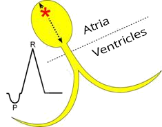
  • In Junctional Rhythm, impulses originate in the AV junction
    • The AV junction consists of the AV node and the His bundle
  • Each impulse then spreads from the AV junction
    • To the atria (producing a negative - retrograde P wave)
    • To the ventricles (producing a QRS complex)
  • Junctional Rhythm has a frequency of 40-60/min.

AV node, AV junction depolarization

Junctional Rhythm - Most Common Causes

Sinus Rhythm and Junctional Rhythm

SA node and sinus rhythm ECG sinus rhythm vs. junctional rhythm

Sinus Rhythm

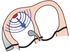
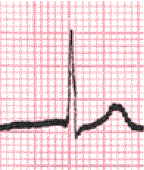
  • Impulses are generated by the SA node with a frequency: 60-100/min.
  • Atrial vector moves toward the AV node (toward lead II)
    • A positive P wave is formed in lead II
  • Then the ventricles are activated
  • The atria are activated first (P wave) and then the ventricles (QRS)


AV node, AV junction ECG junctional rhythm vs. sinus rhythm

Junctional Rhythm

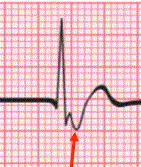
  • Impulses are generated by the AV junction with a frequency of: 40-60/min.
  • From the AV junction, the impulse spreads:
    • to both the atria and the ventricles
  • Ventricular vector moves toward lead II
    • A QRS complex is formed
  • Atrial vector moves toward the SA node
    • Direction is away from lead II
    • A negative - retrograde P wave is formed
  • The ventricles are activated first (QRS) and then the atria (negative P wave)

P Wave and Junctional Rhythm

  • In junctional rhythm, the atria are activated retrogradely
  • P wave is retrograde:
    • Negative in lead II
    • Positive in lead aVR

  • Depending on the timing of depolarization of the ventricles and atria, the P wave may be:
    • Before the QRS (Upper Junctional Rhythm)
    • Within the QRS (Middle Junctional Rhythm)
    • After the QRS (Lower Junctional Rhythm)
atria upwards (P waves) and ventricles downwards (QRS complex)

upper AV nodal junctional rhythm and retrograde P wave middle AV nodal junctional rhythm down AV nodal junctional rhythm and retrograde P wave
Upper Junctional Rhythm
  • P wave before QRS
Middle Junctional Rhythm
  • P wave within QRS
Lower Junctional Rhythm
  • P wave after QRS


upper AV junctional rhythm, retrograde P wave before QRS complex

Upper Junctional Rhythm

  • Impulses originate in the upper part of the AV junction
    • Each impulse simultaneously spreads to both the atria and the ventricles
    • Atria are activated before the ventricles
      • Because the impulse reaches the atria before it reaches the ventricles
  • Shortened PQ interval (< 0.12s)
  • Retrograde P wave is just before the QRS complex


middle AV junctional rhythm, retrograde P wave during QRS complex

Middle Junctional Rhythm

  • Impulses originate in the middle part of the AV junction
    • Each impulse simultaneously spreads to both the atria and the ventricles
    • Atria and ventricles are activated simultaneously
      • The impulse reaches both the atria and the ventricles at the same time
  • Retrograde P wave is hidden within the QRS complex


lower AV junctional rhythm, retrograde P wave after QRS complex

Lower Junctional Rhythm

  • Impulses originate in the lower part of the AV junction
    • Each impulse simultaneously spreads to both the atria and the ventricles
    • Ventricles are activated before the atria
      • Because the impulse reaches the ventricles before it reaches the atria
  • Retrograde P wave is just after the QRS complex

ECG and Junctional Rhythm

  • Retrograde (negative) P wave
    • Negative in the inferior leads (II, III, aVF)
    • Positive in aVR, V1
    • P wave may be before, within, or after the QRS complex
  • Narrow QRS complexes (< 0.12s)
  • Heart rate: 40 - 60/min.
  • Heart rhythm is regular (PP and RR intervals remain unchanged)


junctional AV rhythm atrial and ventricles activation


ECG AV junctional rhythm

Junctional Rhythm

upper AV junctional rhythm, retrograde P wave before QRS complex

Frequency and Junctional Rhythm


ECG junctional av bradycardia, junctional rhythm at a rate of less 40bpm

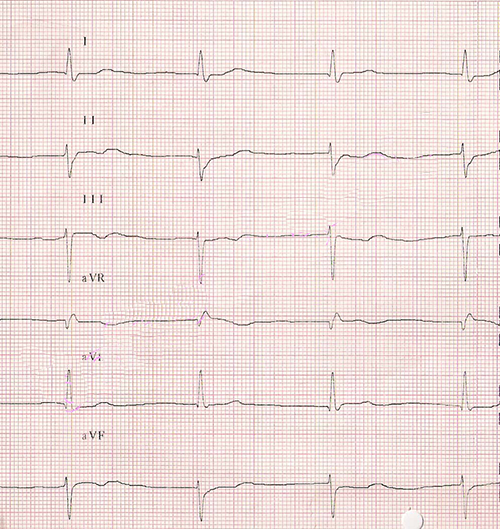
Junctional Bradycardia

  • Frequency: 37/min.
  • Junctional bradycardia has a frequency of < 40/min.
  • P waves are hidden within the QRS complex
    • Impulses are generated by the middle part of the AV junction
middle av junctional rhythm, retrograde P wave during QRS complex


ECG junctional av escape rhythm, junctional rhythm at a rate of 40-60 bpm

Junctional Rhythm

  • Frequency: 45/min.
  • P waves are hidden within the QRS complex
    • Impulses are generated by the middle part of the AV junction
middle av junctional rhythm, retrograde P wave during QRS complex


ECG accelerated av junctional rhythm, junctional rhythm at 60-100 bpm

Accelerated Junctional Rhythm

  • Frequency: 75/min.
  • Retrograde P waves occur after the QRS complexes
    • Impulses are generated by the lower part of the AV junction
lower av junctional rhythm, retrograde P wave after QRS complex


ECG junctional av tachycardia, junctional rhythm more than 100 bpm.

Junctional Tachycardia

  • Frequency: 110/min.
  • Retrograde P waves occur before the QRS complexes
    • Impulses are generated by the upper part of the AV junction

  • Junctional Tachycardia
    • In the AV junction, there is increased automaticity
    • Frequency: 100 - 120/min.
  • AV Nodal Reentry Tachycardia (AVNRT)
    • In the AV junction, there is re-entry
    • Frequency: 120-220/min.
upper av junctional rhythm, retrograde p wave before QRS complex


ECG middle junctional av rhythm, rate 48bpm middle av junctional rhythm, retrograde P wave during QRS complex

Junctional Rhythm


ECG upper av junctional rhythm, retrograde p waves, before qrs

Junctional Rhythm

  • Frequency: 60/min.
  • Retrograde (negative) P waves are before the QRS complexes
    • Impulses are generated by the upper part of the AV junction
  • Narrow QRS complexes (< 0.12s)
upper av junctional rhythm, retrograde p wave before QRS complex


ECG middle av junctional escape rhythm, 45 bpm

Junctional Rhythm

  • Frequency: 45/min.
  • P waves are hidden within the QRS complex
    • Impulses are generated by the middle part of the AV junction
  • Narrow QRS complexes (< 0.12s)
middle av junctional rhythm, retrograde P wave during QRS complex


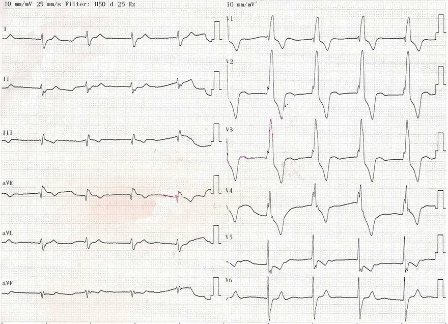
ECG middle av junctional rhythm, right bundle branch block (RBBB), 60 bpm

Junctional Rhythm and Right Bundle Branch Block
  • Frequency: 60/min.
  • P waves are hidden within the QRS complex (not before the QRS - red arrows)
    • Impulses are generated by the middle part of the AV junction
  • Right Bundle Branch Block (RBBB)
    • Wide QRS complexes (> 0.12s)
    • rsR' (V1), right bunny ears are larger
    • Deep S waves (V6)
  • This is a junctional rhythm with aberrant conduction, due to the presence of RBBB
middle av junctional rhythm, retrograde P wave during QRS complex


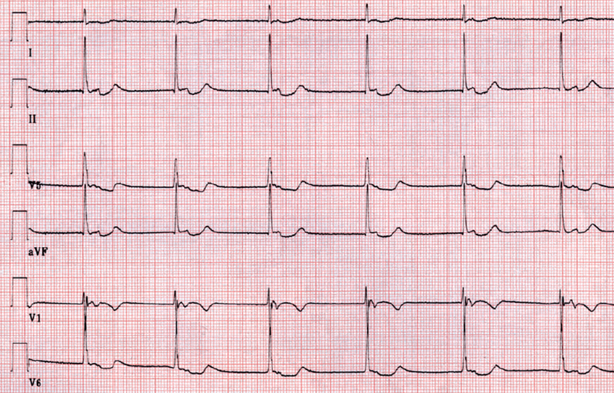
ECG lower av junctional rhythm 60 bpm

Junctional Rhythm

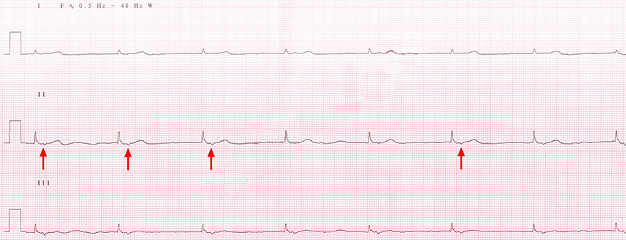
  • Frequency: 60/min.
  • P waves after the QRS complexes (red arrows)
    • Impulses are generated by the lower part of the AV junction
  • Narrow QRS complexes (< 0.12s)
lower av junctional rhythm, retrograde P wave after QRS complex


ECG muddle junctional av bradycardia, 33 bpm middle av junctional rhythm, retrograde P wave during QRS complex

Junctional Bradycardia


ECG isorhythmic av dissociation, sinus rhythm, junctional av rhythm

Junctional Rhythm and Isorhythmic AV Dissociation

  • Sinus Rhythm
    • Frequency: 44/min.
    • Positive P waves AFTER QRS (II lead)
  • Junctional Rhythm
    • Never has positive P waves in the inferior leads (II, III, aVF)
  • At a frequency of 44/min. from the SA node, a junctional rhythm was activated
    • Also with a frequency of 44/min.
  • There are 2 pacemakers in the heart:
    • SA node - sinus rhythm
    • AV junction - junctional rhythm

isorhythmic av dissociation
  • Junctional Rhythm
    • Frequency 44/min.
    • Retrograde P wave of the junctional rhythm does not occur
      • because the P wave originates from the SA node
      • Just before the impulse from the SA node reaches the AV junction, the AV junction generates its own impulse.
      • Then, just above the AV junction, the 2 impulses meet and cancel each other out during the (refractory period)
  • Isorhythmic AV Dissociation
    • It is the independence of the atria (P waves) from the ventricles (QRS) with the same frequency


Sources

  • ECG from Basics to Essentials Step by Step
  • litfl.com
  • ecgwaves.com
  • metealpaslan.com
  • medmastery.com
  • uptodate.com
  • ecgpedia.org
  • wikipedia.org
  • Strong Medicine
  • Understanding Pacemakers