ECGbook.com

Making Medical Education Free for All

ECGbook.com

Making Medical Education Free for All

Home /

Persistent juvenile T-wave pattern

Persistent juvenile T wave

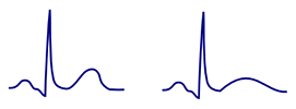
Normal T Wave

ECG normal positive T wave

Concordant T Wave


ECG abnormal disconcordance T wave, positive QRS, negative inverted T wave

Disconcordant T Wave


  • The T wave is the most variable wave on the ECG
  • Is concordant (has the same polarity as the QRS)
    • Positive QRS - positive T wave
    • Negative QRS - negative T wave
  • Is positive (I, II, V3-V6)
    • Can be isolated negative (III, aVL)
  • Is negative (aVR)
  • Amplitude
  • For ECG diagnostics, the dynamic nature of the T wave is important
    • If the T wave is consistently the same - it often does not indicate a serious condition
    • If the T wave changes - it often indicates a serious condition (ischemia)

ECG and Persistent Juvenile T Waves

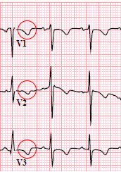
ECG (V1-V3) persistent juvenile T wave pattern (PJTWP), inverted T wave

Differential Diagnosis


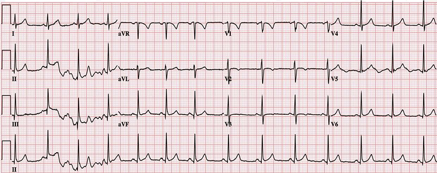
ECG (V1-V4) persistent juvenile T wave pattern, african-american female

Persistent Juvenile T Waves


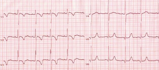
ECG (V1-V3) persistent juvenile T wave pattern, shallow asymmetric morphology inversions T waves (V1-V3), no significant ST segment

Persistent Juvenile T Waves

  • It was a 5-year-old healthy child without structural heart disease
  • Inverted T waves (V1-V2)
    • Depth < 3mm
  • The T waves did not change in repeated EKG checks


ECG (V1-V3) persistent juvenile T wave pattern (PJTWI), Benign T wave inversion (BTWI), assymetric T wave inversion

Persistent Juvenile T Waves


ECG (V1-V3) persistent juvenile T wave pattern (PJTWI)

Persistent Juvenile T Waves

  • It was a 20-year-old healthy woman without structural heart disease
  • Inverted T waves (V1-V3)
    • Depth < 3mm (sometimes the depth can exceed 3mm)
  • The T waves did not change in repeated EKG checks


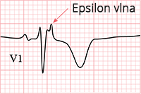
ECG epsilon wave, inverted T wave

ECG (V1-V3) arrhythmogenic right ventricular hypertrophy, epsilon wave, inverted T wave

Arrhythmogenic Right Ventricular Dysplasia


Heart stenosis: Circumflex branch of left coronary artery (LCx), Anterior interventricular branch of left coronary artery (LAD)
ECG ischemic inverted T waves, unstable angina pectoris, stenosis LAD, LCx

Inverted Ischemic T Waves and Unstable Angina Pectoris

  • The patient had unstable angina pectoris due to stenosis of the LAD and LCx
    • ST depression (V4-V6)
    • Inverted T waves (V2-V6)
    • After stent placement (recanalization), the T waves normalized later
  • For acute subendocardial ischemia, the following are characteristic
    • ST depression and Inverted T waves (at least in 2 contiguous leads)
  • Persistent juvenile T waves are only present in leads (V1-V3)


Sources

  • ECG from Basics to Essentials Step by Step
  • litfl.com
  • ecgwaves.com
  • metealpaslan.com
  • medmastery.com
  • uptodate.com
  • ecgpedia.org
  • wikipedia.org
  • Strong Medicine
  • Understanding Pacemakers


šípka späť

Persistent juvenile T-wave pattern

Persistent juvenile T wave

Normal T Wave

  • The T wave is the most variable wave on the ECG
  • Is concordant (has the same polarity as the QRS)
    • Positive QRS - positive T wave
    • Negative QRS - negative T wave
  • Is positive (I, II, V3-V6)
    • Can be isolated negative (III, aVL)
  • Is negative (aVR)
  • Amplitude
  • For ECG diagnostics, the dynamic nature of the T wave is important
    • If the T wave is consistently the same - it often does not indicate a serious condition
    • If the T wave changes - it often indicates a serious condition (ischemia)
ECG normal positive T wave

Concordant T Wave


ECG abnormal disconcordance T wave, positive QRS, negative inverted T wave

Disconcordant T Wave


ECG and Persistent Juvenile T Waves

  • Occur primarily in African-American women under 30 years old
    • Present from childhood and persist into adulthood (do not change)
  • Likely caused by hemodynamic right ventricular overload
    • during intrauterine development
  • Common in athletes
  • Patients do not have structural heart damage

  • ECG and Persistent Juvenile T Waves
    • Seen in leads V1-V3
      • In children under 12 years, they can also be present in V4
    • Are asymmetric inverted T waves
    • Are deep < 3mm (most common, but not a rule)


ECG (V1-V3) persistent juvenile T wave pattern (PJTWP), inverted T wave

Differential Diagnosis


ECG (V1-V4) persistent juvenile T wave pattern, african-american female

Persistent Juvenile T Waves


ECG (V1-V3) persistent juvenile T wave pattern, shallow asymmetric morphology inversions T waves (V1-V3), no significant ST segment

Persistent Juvenile T Waves

  • It was a 5-year-old healthy child without structural heart disease
  • Inverted T waves (V1-V2)
    • Depth < 3mm
  • The T waves did not change in repeated EKG checks


ECG (V1-V3) persistent juvenile T wave pattern (PJTWI), Benign T wave inversion (BTWI), assymetric T wave inversion

Persistent Juvenile T Waves


ECG (V1-V3) persistent juvenile T wave pattern (PJTWI)

Persistent Juvenile T Waves

  • It was a 20-year-old healthy woman without structural heart disease
  • Inverted T waves (V1-V3)
    • Depth < 3mm (sometimes the depth can exceed 3mm)
  • The T waves did not change in repeated EKG checks


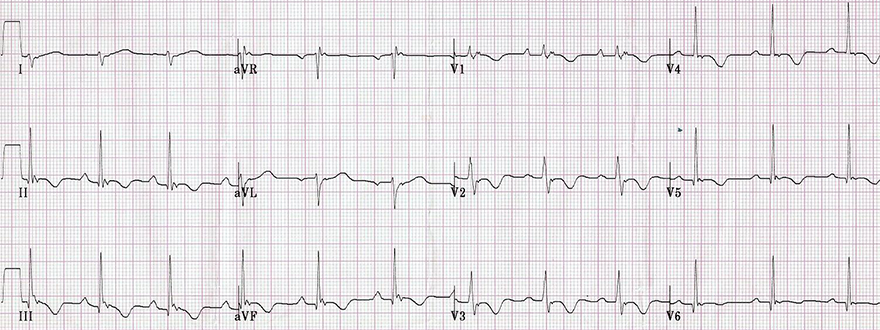
ECG (V1-V3) arrhythmogenic right ventricular hypertrophy, epsilon wave, inverted T wave

Arrhythmogenic Right Ventricular Dysplasia
ECG epsilon wave, inverted T wave


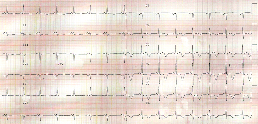
ECG ischemic inverted T waves, unstable angina pectoris, stenosis LAD, LCx

Inverted Ischemic T Waves and Unstable Angina Pectoris

  • The patient had unstable angina pectoris due to stenosis of the LAD and LCx
    • ST depression (V4-V6)
    • Inverted T waves (V2-V6)
    • After stent placement (recanalization), the T waves normalized later
  • For acute subendocardial ischemia, the following are characteristic
    • ST depression and Inverted T waves (at least in 2 contiguous leads)
  • Persistent juvenile T waves are only present in leads (V1-V3)
Heart stenosis: Circumflex branch of left coronary artery (LCx), Anterior interventricular branch of left coronary artery (LAD)


Sources

  • ECG from Basics to Essentials Step by Step
  • litfl.com
  • ecgwaves.com
  • metealpaslan.com
  • medmastery.com
  • uptodate.com
  • ecgpedia.org
  • wikipedia.org
  • Strong Medicine
  • Understanding Pacemakers