|
|
ECGbook.com Making Medical Education Free for All |
Upload ECG for Interpretation |
|
|
ECGbook.com Making Medical Education Free for All |
Upload ECG for Interpretation |
|
|
ECGbook.com Making Medical Education Free for All |
Home /
Left Anterior Hemiblock (LAHB), Left Anterior Fascicular Block

Left Anterior Fascicular Block (Quick Diagnosis)


Differential Diagnosis Using V1-V2


Left Anterior Fascicular Block
Left Anterior Fascicular Block

Trifascicular Block (Left Anterior Fascicular Block + AV Block of 1st Degree + RBBB)
Sources
Home /
Left Anterior Hemiblock (LAHB), Left Anterior Fascicular Block
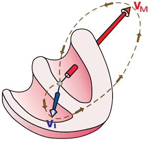
Left Bundle Branch
|
|
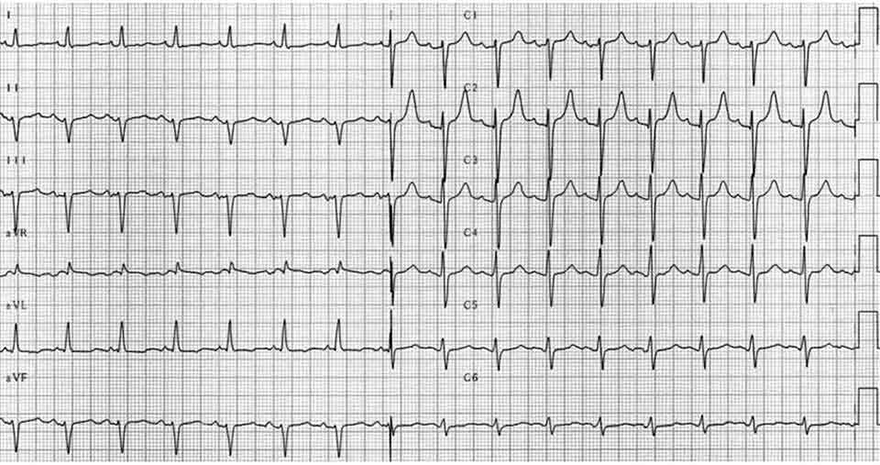
Left Anterior Fascicular Block
|
|
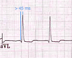
Left Anterior Fascicular Block (Quick Diagnosis)
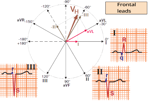
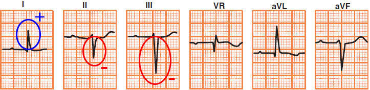
ECG and Left Anterior Fascicular Block
|
|
Differential Diagnosis
|
|
|
Differential Diagnosis Using V1-V2
|
|
|
Left Anterior Fascicular Block
|
|
|
Left Anterior Fascicular Block
|
|
Trifascicular Block (Left Anterior Fascicular Block + AV Block of 1st Degree + RBBB)
Sources