|
|
ECGbook.com Making Medical Education Free for All |
Upload ECG for Interpretation |
|
|
ECGbook.com Making Medical Education Free for All |
Upload ECG for Interpretation |
|
|
ECGbook.com Making Medical Education Free for All |


Acute Posterior-Lateral STEMI

Acute Lateral STEMI


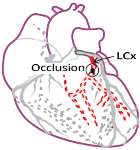
Coronary Angiography


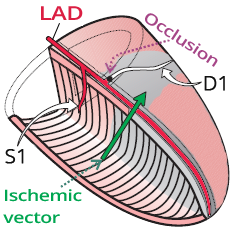
Acute Antero-lateral STEMI


Acute Antero-lateral STEMI
Acute Isolated High Lateral STEMI
Acute Isolated High Lateral STEMI

Subacute Isolated High Lateral STEMI

Acute Inferior-Posterior-Lateral STEMI
Sources
|
|
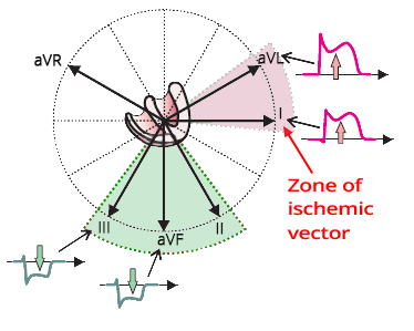
Lateral Infarction
|
|
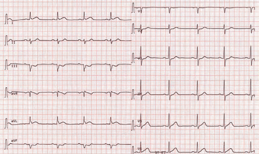
ECG and Lateral STEMI
|
|
|
|
Antero-lateral STEMI
|
|
Infero-posterior-lateral STEMI
|
|
|
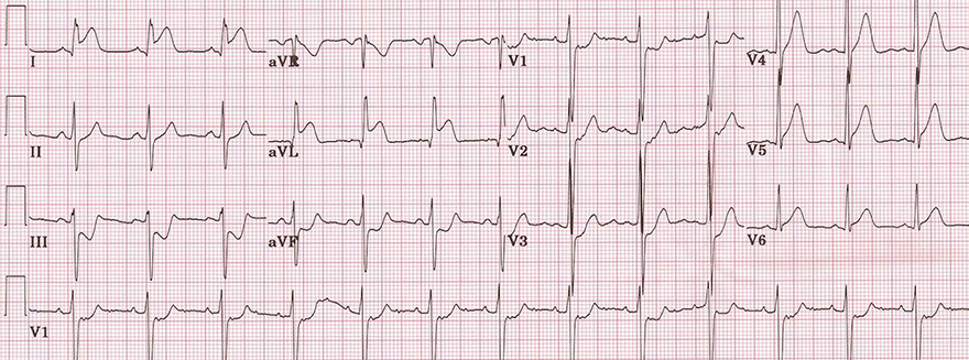
Acute Posterior-Lateral STEMI
|
|
|
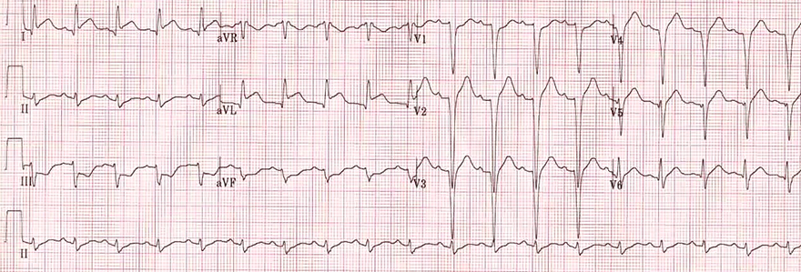
Acute Lateral STEMI
|
|
Coronary Angiography
|
Acute Antero-lateral STEMI
|
|
|
Acute Antero-lateral STEMI
|
|
|
Acute Isolated High Lateral STEMI
|
|
|
Acute Isolated High Lateral STEMI
|
|
|
Subacute Isolated High Lateral STEMI
|
|
|
Acute Inferior-Posterior-Lateral STEMI
|
|
Sources