ECGbook.com

Making Medical Education Free for All

ECGbook.com

Making Medical Education Free for All

Home /

Left Posterior Fascicular Block

Left Posterior Hemiblock (LPHB), Left Posterior Fascicular Block

Left Bundle Branch

Heart conduction system, left bundle branch, left posterior fascicle
  • Left Bundle Branch (LBB) divides after a short course into:
    • Left Anterior Fascicle
      • Runs anteriorly and laterally
      • Activates the anterior and lateral part of the left ventricle
    • Left Posterior Fascicle
      • Runs posteriorly and inferiorly
      • Activates the posterior and inferior part of the left ventricle

    • The Left Anterior Fascicle is thinner than the Left Posterior Fascicle

Left Posterior Fascicular Block

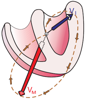
Left posterior Fascicular Block electrical vectors

Electrical Vector and Left Posterior Fascicular Block

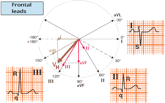
Left posterior Fascicular Block, frontal leads, projections of the main vector

Left Anterior Fascicular Block (Quick Diagnosis)
  • Left Posterior Fascicular Block is diagnosed using limb leads (I, II, III)
  • The main vector (VH) points in the frontal plane down to the right
    • Lead I is negative
    • Lead III is positive
      • R wave in III > aVF > II

ECG and Left Posterior Fascicular Block

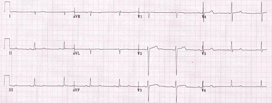
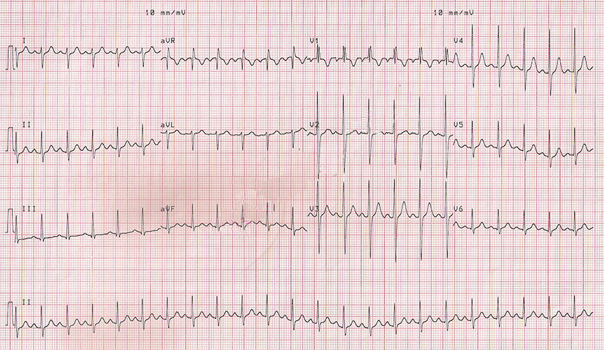
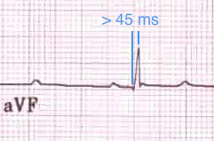
ECG Left Posterior Fascicular Block (LPFB), Prolonged R wave peak time in aVF 45ms, negative QRS I lead, positive QRS III lead ECG left posterior Fascicular Block, Right axis deviation, QRS duration normal or slightly prolonged

Differential Diagnosis

Right axis deviation, diagnosis left posterior Fascicular Block (LPH) of exclusion

Right axis deviation, left posterior Fascicular Block
ECG Left Posterior Fascicular Block (LPFB), right axis deviation

Left Posterior Fascicular Block


Right axis deviation, left posterior Fascicular Block
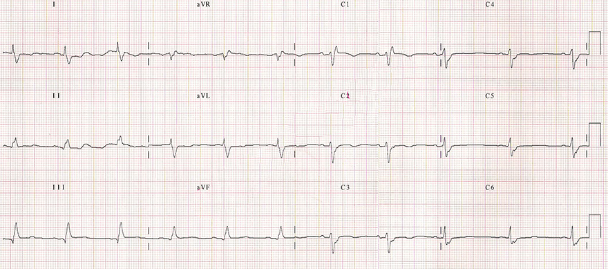
ECG left posterior Fascicular Block, right axis deviation, narrow QRS, prolonged R wave peak aVF

Left Posterior Fascicular Block

  • Narrow QRS complexes (< 0.12s)
  • Right Axis Deviation (>90°)
    • Negative QRS in lead I
    • Positive QRS in lead III
  • All causes of right axis deviation must be excluded in the patient
  • LPFB almost always occurs together with RBBB (Bifascicular Block)


Right axis deviation, left posterior Fascicular Block
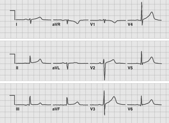
ECG left posterior fascicular block, right axis deviation, deep S waves (I, aVL), tall R waves (III, II, aVF)

Left Posterior Fascicular Block


Right axis deviation, left posterior Fascicular Block
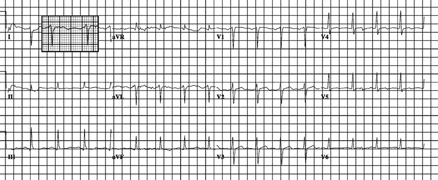
ECG left posterior fascicular block (LPFB), deep S waves (I, aVL), tall R waves (III, II, aVF)

Left Posterior Fascicular Block

  • Narrow QRS complexes (< 0.12s)
  • Right Axis Deviation (>90°)
    • Negative QRS in lead I
    • Positive QRS in lead III
  • All causes of right axis deviation must be excluded in the patient
  • LPFB almost always occurs together with RBBB (Bifascicular Block)


Bifascicular block, left posterior fascicular block (LPFB), incomplete right bundle branch block (iRBBB)
ECG bifascicular block, left posterior fascicular block (LPFB), incomplete right bundle branch block (iRBBB)

Bifascicular Block (LPFB + iRBBB)


Bifascicular block, left posterior fascicular block (LPFB), complete right bundle branch block (RBBB)
ECG bifascicular block (LPFB + RBBB)

Bifascicular Block (LPFB + RBBB)


Bifascicular block, left posterior fascicular block (LPFB), complete right bundle branch block (RBBB)
ECG bifascicular block, Extreme (right) axis deviation, left posterior fascicular block, right bundle branch block (RBBB)

Bifascicular Block (LPFB + RBBB)


Sources

  • ECG from Basics to Essentials Step by Step
  • litfl.com
  • ecgwaves.com
  • metealpaslan.com
  • medmastery.com
  • uptodate.com
  • ecgpedia.org
  • wikipedia.org
  • Strong Medicine
  • Understanding Pacemakers


šípka späť

Left Posterior Fascicular Block

Left Posterior Hemiblock (LPFB), Left Posterior Fascicular Block

Left Bundle Branch

  • Left Bundle Branch (LBB) divides after a short course into:
    • Left Anterior Fascicle
      • Runs anteriorly and laterally
      • Activates the anterior and lateral part of the left ventricle
    • Left Posterior Fascicle
      • Runs posteriorly and inferiorly
      • Activates the posterior and inferior part of the left ventricle

    • The Left Anterior Fascicle is thinner than the Left Posterior Fascicle

Heart conduction system, left bundle branch, left posterior fascicle

Left Posterior Fascicular Block

  • The Left Posterior Fascicle is thick and has dual blood supply
  • Left Posterior Fascicular Block
  • In Left Posterior Fascicular Block
    • the lateral part of the left ventricle is activated first
      • via the anterior fascicle
      • A small initial vector (VI) is formed
    • Then the inferior part of the ventricles is activated
      • Via the myocardium, because the left posterior fascicle is blocked
      • The main cardiac vector (VH) is formed and rotates to the right

Left posterior Fascicular Block electrical vectors

Electrical Vector and Left Posterior Fascicular Block

Left posterior Fascicular Block, frontal leads, projections of the main vector

Left Anterior Fascicular Block (Quick Diagnosis)

  • Left Posterior Fascicular Block is diagnosed using limb leads (I, II, III)
  • The main vector (VH) points in the frontal plane down to the right
    • Lead I is negative
    • Lead III is positive
      • R wave in III > aVF > II

ECG and Left Posterior Fascicular Block

  • Narrow QRS complex < 0.12s
    • QRS is widened by approximately 20ms, but does not exceed 0.12s
  • Right Axis Deviation > 90º
    • Lead I is negative
    • Lead III is positive
      • R wave in III > aVF > II
  • Prolonged R wave peak time in aVF > 45ms
    • From the start of the QRS complex to the peak of the R wave is more than 45ms
    • Because activation of the inferior part of the left ventricle is prolonged
      • The part that should be activated through the blocked posterior fascicle

  • LPFB almost always occurs together with RBBB (Bifascicular Block)

ECG Left Posterior Fascicular Block (LPFB), Prolonged R wave peak time in aVF 45ms, negative QRS I lead, positive QRS III lead

ECG left posterior Fascicular Block, Right axis deviation, QRS duration normal or slightly prolonged

Differential Diagnosis


Right axis deviation, diagnosis left posterior Fascicular Block (LPH) of exclusion


ECG Left Posterior Fascicular Block (LPFB), right axis deviation

Left Posterior Fascicular Block

  • Narrow QRS complexes (< 0.12s)
  • Right Axis Deviation (>90°)
    • Negative QRS in lead I
    • Positive QRS in lead III
  • All causes of right axis deviation must be excluded in the patient
  • LPFB almost always occurs together with RBBB (Bifascicular Block)
Right axis deviation, left posterior Fascicular Block


ECG left posterior Fascicular Block, right axis deviation, narrow QRS, prolonged R wave peak aVF

Left Posterior Fascicular Block

  • Narrow QRS complexes (< 0.12s)
  • Right Axis Deviation (>90°)
    • Negative QRS in lead I
    • Positive QRS in lead III
  • All causes of right axis deviation must be excluded in the patient
  • LPFB almost always occurs together with RBBB (Bifascicular Block)
Right axis deviation, left posterior Fascicular Block


ECG left posterior fascicular block, right axis deviation, deep S waves (I, aVL), tall R waves (III, II, aVF)

Left Posterior Fascicular Block
  • Narrow QRS complexes (< 0.12s)
  • Right Axis Deviation (>90°)
    • Negative QRS in lead I
    • Positive QRS in lead III
  • All causes of right axis deviation must be excluded in the patient
  • LPFB almost always occurs together with RBBB (Bifascicular Block)
Right axis deviation, left posterior Fascicular Block


ECG left posterior fascicular block (LPFB), deep S waves (I, aVL), tall R waves (III, II, aVF)

Left Posterior Fascicular Block

  • Narrow QRS complexes (< 0.12s)
  • Right Axis Deviation (>90°)
    • Negative QRS in lead I
    • Positive QRS in lead III
  • All causes of right axis deviation must be excluded in the patient
  • LPFB almost always occurs together with RBBB (Bifascicular Block)
Right axis deviation, left posterior Fascicular Block


ECG bifascicular block, left posterior fascicular block (LPFB), incomplete right bundle branch block (iRBBB)

Bifascicular Block (LPFB + iRBBB)


Bifascicular block, left posterior fascicular block (LPFB), incomplete right bundle branch block (iRBBB)


ECG bifascicular block (LPFB + RBBB)

Bifascicular Block (LPFB + RBBB)
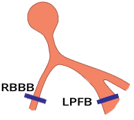

Bifascicular block, left posterior fascicular block (LPFB), complete right bundle branch block (RBBB)

ECG bifascicular block, Extreme (right) axis deviation, left posterior fascicular block, right bundle branch block (RBBB)

Bifascicular Block (LPFB + RBBB)


Bifascicular block, left posterior fascicular block (LPFB), complete right bundle branch block (RBBB)


Sources

  • ECG from Basics to Essentials Step by Step
  • litfl.com
  • ecgwaves.com
  • metealpaslan.com
  • medmastery.com
  • uptodate.com
  • ecgpedia.org
  • wikipedia.org
  • Strong Medicine
  • Understanding Pacemakers