ECGbook.com

Making Medical Education Free for All

ECGbook.com

Making Medical Education Free for All

Home /

Aneurysm of the Left Ventricle

Left ventricular aneurysm

STEMI and the Left Ventricle

Normal left ventricular wall
  • STEMI is a transmural myocardial infarction
    • ST elevations occur, which have dynamics and typically resolve within 2 weeks
    • Myocardial wall necrosis occurs (electrical window)
    • The necrotic wall is weakened; if it dilates, it may form an aneurysm
    • STEMI is the most common cause of left ventricular aneurysm
      • Right ventricular aneurysm is rare because the right ventricle has low pressure

Aneurysm of the Left Ventricle

Left ventricular aneurysm after STEMI infarction

ECG and Left Ventricular Aneurysm

ECG criteria left ventricular aneurysm after STEMI, persistent ST elevation

Dynamics of ST Elevations in STEMI


ECG evolution STEMI infarction, persistent ST elevation

Classification of STEMI by Stage
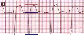

T/QRS Ratio (V1-4)

  • Used for differential diagnosis:
    • Left ventricular aneurysm vs. STEMI
  • In practice, it is rarely used (has only a rough indication)

  • Left ventricular aneurysm has T/QRS < 0.36 in all precordial leads (V1-V6)
  • Acute STEMI has T/QRS > 0.36 in at least one precordial lead

ECG (V1) T/QRS ratio, distinguishes ventricular aneurysm from anterior STEMI myocardial infarction
T=1, QRS=11, T/QRS = 0.09

ECG (V2) T/QRS ratio, distinguishes ventricular aneurysm from anterior STEMI myocardial infarction
T=1.5, QRS=13, T/QRS = 0.11

ECG (V3) T/QRS ratio, distinguishes ventricular aneurysm from anterior STEMI myocardial infarction
T=6, QRS=18, T/QRS = 0.33

ECG (V4) T/QRS ratio, distinguishes ventricular aneurysm from anterior STEMI myocardial infarction
T=5, QRS=13, T/QRS = 0.38


Acute STEMI Infarction

  • All 4 leads are from the same patient
  • The patient had chest pain, and the older ECG did not show these changes
  • T/QRS Ratio = 0.38 (V4) is > 0.36 (indicating acute STEMI)


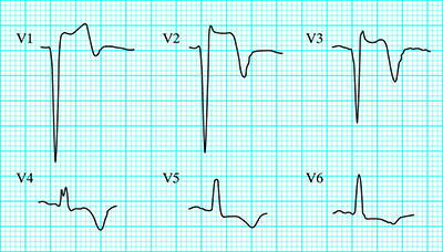
Left ventricular aneurysm of anterior wall
ECG anterior Left Ventricular Aneurysm, Minimal ST elevation (V1-V3), deep Q waves and T waves inversion

Anterior Wall Aneurysm


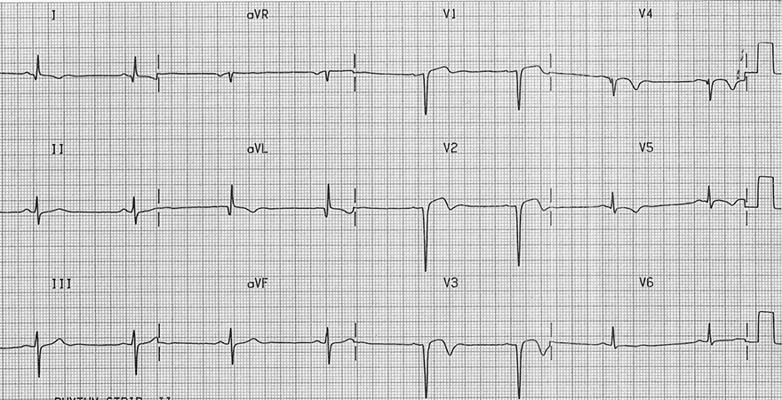
Left ventricular aneurysm of inferior wall, LV aneurysm morphology
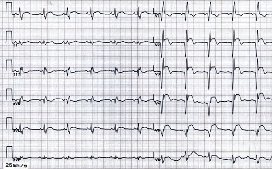
ECG Inferior Left Ventricular Aneurysm, old inferior STEMI, persistent ST elevation

Inferior Wall Aneurysm


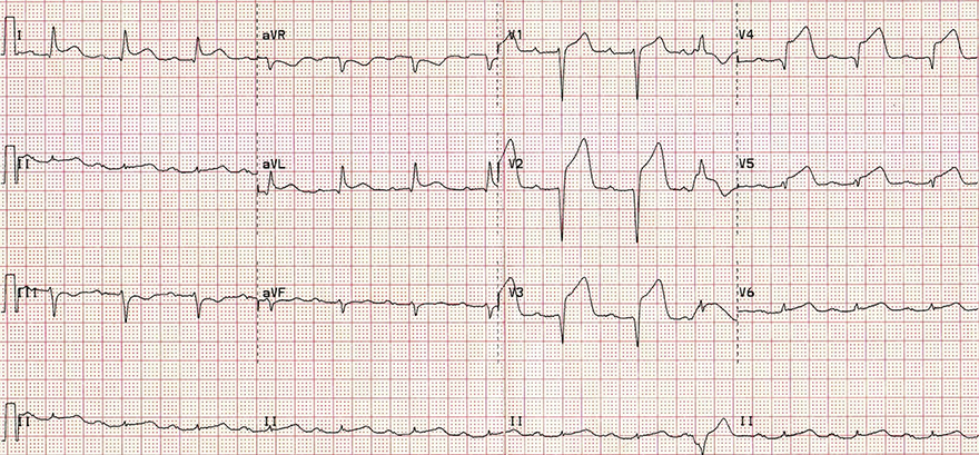
Anterior STEMI infarction
ECG acute anterior STEMI infarction, anterior aneurysm formation

Acute Anterior STEMI


Acute inferior STEMI infarction, inferior wall aneurysm formation
ECG acute inferior STEMI, inferior wall aneurysm formation

Acute Inferior STEMI

  • The patient experienced chest pain and was sweaty
  • Acute Inferior STEMI
    • ST elevations (II, III, aVF)
      • ST elevation III > II (Occlusion is distal in the right coronary artery)
    • Reciprocal ST depression (aVL)
    • Pathological Q waves (III, aVF)
  • In 5% of patients after an inferior STEMI, an aneurysm will develop
    • In the case of an aneurysm: ST elevations will not resolve within 2 weeks and will be reduced


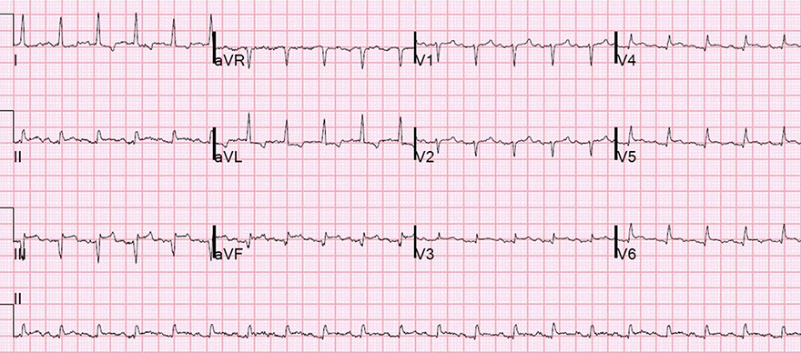
Left ventricular aneurysm after anterior STEMI infarction
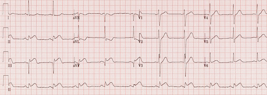
ECG old anterior myocardial infarction, persistent ST elevation, left ventricular aneurysm

Anterior Wall Aneurysm


Left ventricular aneurysm after anterior STEMI infarction
ECG old anterior myocardial STEMI infarction, persistent ST elevation, left ventricular aneurysm

Anterior Wall Aneurysm


Left ventricular aneurysm after anterior STEMI infarction
ECG persistent ST elevation, left ventricular aneurysm, old anterior myocardial STEMI infarction

Anterior Wall Aneurysm


Sources

  • ECG from Basics to Essentials Step by Step
  • litfl.com
  • ecgwaves.com
  • metealpaslan.com
  • medmastery.com
  • uptodate.com
  • ecgpedia.org
  • wikipedia.org
  • Strong Medicine
  • Understanding Pacemakers


šípka späť

Aneurysm of the Left Ventricle

Left ventricular aneurysm

STEMI and the Left Ventricle

  • STEMI is a transmural myocardial infarction
    • ST elevations occur, which have dynamics and typically resolve within 2 weeks
    • Myocardial wall necrosis occurs (electrical window)
    • The necrotic wall is weakened; if it dilates, it may form an aneurysm
    • STEMI is the most common cause of left ventricular aneurysm
      • Right ventricular aneurysm is rare because the right ventricle has low pressure

Normal left ventricular wall

Aneurysm of the Left Ventricle

  • Causes of left ventricular aneurysm:
  • Risks of left ventricular aneurysm
    • Ventricular tachycardia (left ventricle is structurally damaged)
    • Aneurysm rupture (patient can bleed out immediately)
    • Embolization to the systemic circulation (thrombus can form in the aneurysm)


Left ventricular aneurysm after STEMI infarction

ECG and Left Ventricular Aneurysm

ECG criteria left ventricular aneurysm after STEMI, persistent ST elevation

Dynamics of ST Elevations in STEMI


ECG evolution STEMI infarction, persistent ST elevation

Classification of STEMI by Stage


T/QRS Ratio (V1-4)

  • Used for differential diagnosis:
    • Left ventricular aneurysm vs. STEMI
  • In practice, it is rarely used (has only a rough indication)

  • Left ventricular aneurysm has T/QRS < 0.36 in all precordial leads (V1-V6)
  • Acute STEMI has T/QRS > 0.36 in at least one precordial lead

ECG (V1) T/QRS ratio, distinguishes ventricular aneurysm from anterior STEMI myocardial infarction
T=1, QRS=11, T/QRS = 0.09

ECG (V2) T/QRS ratio, distinguishes ventricular aneurysm from anterior STEMI myocardial infarction
T=1.5, QRS=13, T/QRS = 0.11
ECG (V3) T/QRS ratio, distinguishes ventricular aneurysm from anterior STEMI myocardial infarction
T=6, QRS=18, T/QRS = 0.33

ECG (V4) T/QRS ratio, distinguishes ventricular aneurysm from anterior STEMI myocardial infarction
T=5, QRS=13, T/QRS = 0.38

Acute STEMI Infarction

  • All 4 leads are from the same patient
  • The patient had chest pain, and the older ECG did not show these changes
  • T/QRS Ratio = 0.38 (V4) is > 0.36 (indicating acute STEMI)


ECG anterior Left Ventricular Aneurysm, Minimal ST elevation (V1-V3), deep Q waves and T waves inversion

Anterior Wall Aneurysm

Left ventricular aneurysm of anterior wall


ECG Inferior Left Ventricular Aneurysm, old inferior STEMI, persistent ST elevation

Inferior Wall Aneurysm

Left ventricular aneurysm of inferior wall, LV aneurysm morphology


ECG acute anterior STEMI infarction, anterior aneurysm formation

Acute Anterior STEMI

  • The patient experienced chest pain and was sweaty
  • Acute Anterior STEMI
    • ST elevations (V2-5, I, aVL)
    • Reciprocal ST depression (III)
    • Pathological Q waves (V2-3)
  • In 60% of patients after an anterior STEMI, an aneurysm will develop
    • In the case of an aneurysm: ST elevations will not resolve within 2 weeks and will be reduced
Anterior STEMI infarction


ECG acute inferior STEMI, inferior wall aneurysm formation

Acute Inferior STEMI

  • The patient experienced chest pain and was sweaty
  • Acute Inferior STEMI
    • ST elevations (II, III, aVF)
      • ST elevation III > II (Occlusion is distal in the right coronary artery)
    • Reciprocal ST depression (aVL)
    • Pathological Q waves (III, aVF)
  • In 5% of patients after an inferior STEMI, an aneurysm will develop
    • In the case of an aneurysm: ST elevations will not resolve within 2 weeks and will be reduced
Acute inferior STEMI infarction, inferior wall aneurysm formation


ECG old anterior myocardial infarction, persistent ST elevation, left ventricular aneurysm

Anterior Wall Aneurysm

Left ventricular aneurysm after anterior STEMI infarction


ECG old anterior myocardial STEMI infarction, persistent ST elevation, left ventricular aneurysm

Anterior Wall Aneurysm

Left ventricular aneurysm after anterior STEMI infarction


ECG persistent ST elevation, left ventricular aneurysm, old anterior myocardial STEMI infarction

Anterior Wall Aneurysm

Left ventricular aneurysm after anterior STEMI infarction


Sources

  • ECG from Basics to Essentials Step by Step
  • litfl.com
  • ecgwaves.com
  • metealpaslan.com
  • medmastery.com
  • uptodate.com
  • ecgpedia.org
  • wikipedia.org
  • Strong Medicine
  • Understanding Pacemakers