ECGbook.com

Making Medical Education Free for All

ECGbook.com

Making Medical Education Free for All

Home /

Left Ventricular Hypertrophy

Left ventricular hypertrophy

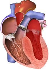
Left Ventricle

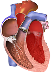
Normal left ventricle thickness
  • The left ventricle has a physiological thickness of 10-12mm
  • Hemodynamic overload of the ventricle (volume or pressure) causes remodeling of the ventricle
    • Left ventricular hypertrophy
      • It is wall thickening > 12mm
      • The wall of the left ventricle has a physiological thickness of 10-12mm
    • Right ventricular hypertrophy
      • It is wall thickening > 5mm
      • The wall of the right ventricle has a physiological thickness of 3-4mm
  • According to hemodynamic overload, we recognize 2 types of hypertrophy:
    • Concentric hypertrophy
    • Excentric hypertrophy

Concentric Left Ventricular Hypertrophy

left ventricle concentric hypertrophy

Eccentric Left Ventricular Hypertrophy

left ventricle eccentric hypertrophy
  • The ventricle is overloaded by volume (increased preload)
    • It becomes filled with blood during diastole
    • During systole, it is overloaded by pressure (pumps a larger volume of blood)
  • Dilation and hypertrophy of the ventricular cavity occurs
    • With chronic volume overload, only dilation occurs
  • Eccentric left ventricular hypertrophy, causes:
    • Aortic insufficiency
  • Eccentric right ventricular hypertrophy, causes:
    • Pulmonary insufficiency

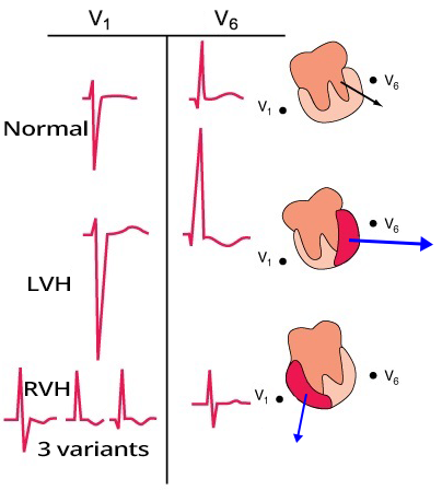
Main Ventricular Vector and Ventricular Hypertrophy

ECG pattern features V1, V6, left ventricular hypertrophy, right ventricular hypertrophy

Left Ventricular Hypertrophy

  • ECG cannot differentiate between concentric and eccentric hypertrophy
  • ECG in the diagnosis of left ventricular hypertrophy has:
    • Low sensitivity (20-50%)
      • Many patients with hypertrophy have a normal ECG (false negatives)
    • High specificity (90%)
      • If a patient has ECG signs of hypertrophy, then hypertrophy is very likely
  • Diagnosis of hypertrophy is performed using echocardiography
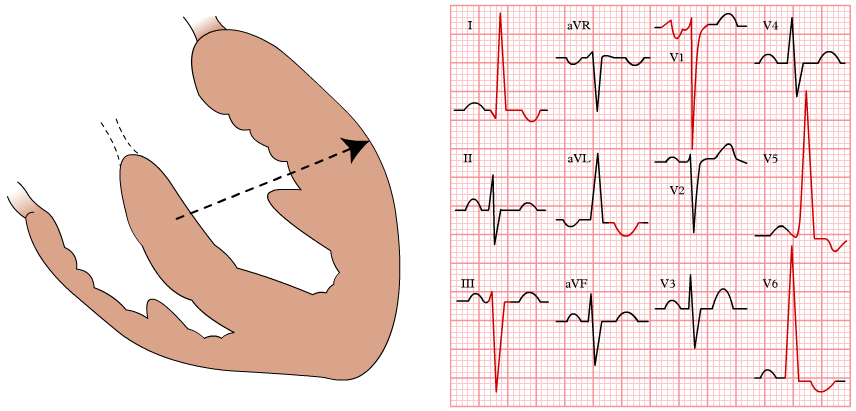
  • The principle of ECG diagnosis of LVH is that the cardiac vector rotates:
    • Toward lateral leads (I, aVL, V5-V6)
    • Away from right-sided leads (aVR, III, V1-V2)

ECG and Left Ventricular Hypertrophy


Sokolow Index

  • S(V1/2) + R(V5/6) ≥ 35mm
  • It is the most commonly used criterion

  • Cornell Index
    • More sensitive than the Sokolow index
    • Less commonly remembered (often not used)
    • Men: R(aVL) + S(V3) > 28mm
    • Women: R(aVL) + S(V3) > 20mm
  • Cornell Product Duration
    • Has the highest sensitivity
    • Less commonly remembered (often not used)
    • Cornell Index (mm) x QRS Duration (ms) > 2440 mm.ms
ECG, voltage criteria, Sokolow Lyon criteria, index, left ventricular hypertrophy

Lewis Index

ECG, voltage criteria, Lewis index, left ventricular hypertrophy

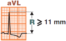
aVL(R) ≥ 11mm

ECG, voltage criteria, left ventricular hypertrophy, R wave in aVL 11mm

aVL(R) ≥ 13mm and III(S) ≥ 15mm

ECG, voltage criteria, left ventricular hypertrophy, aVL(R) 13mm and III(S) 15mm

Prolonged R Wave Peak Time (V6) ≥ 50ms

ECG R Wave Peak Time (Intrinsicoid Deflection), left ventricular hypertrophy
  • The time from the start of the QRS complex to the peak of the R wave is referred to as:
    • R Wave Peak Time (Intrinsicoid Deflection)

  • Prolonged R Wave Peak Time (V6) ≥ 50ms
    • Is seen in left ventricular hypertrophy

  • In hypertrophy, the time for wall activation is prolonged
  • Thus, the distance between:


ECG left ventricular hypertrophy, voltage criteria, sokolov lyon ciretir, lewis index, r wave in aVL, increased r wave peak time

Left Ventricular Hypertrophy


Left Ventricular Overload and Concentric Hypertrophy of the LV

Left ventricular strain pattern

  • Only concentric hypertrophy of the LV (pressure overload) is created
    • Not eccentric hypertrophy of the LV (volume overload)

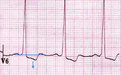
  • Descending ST depressions in lateral leads (I, aVL, V5-6)
ECG Left ventricular strain pattern, ST depression, T wave inversion in lateral leads


ECG left ventricular hypertrophy, voltage criteria, increased LV voltages, r wave peak time, LV strain pattern, ST elevation, left axis deviation

Left Ventricular Hypertrophy


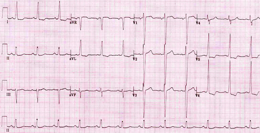
ECG left ventricular hypertrophy (LVH), voltage criteria, increased LV voltages, r wave peak time, LV strain pattern, ST elevation, left axis deviation

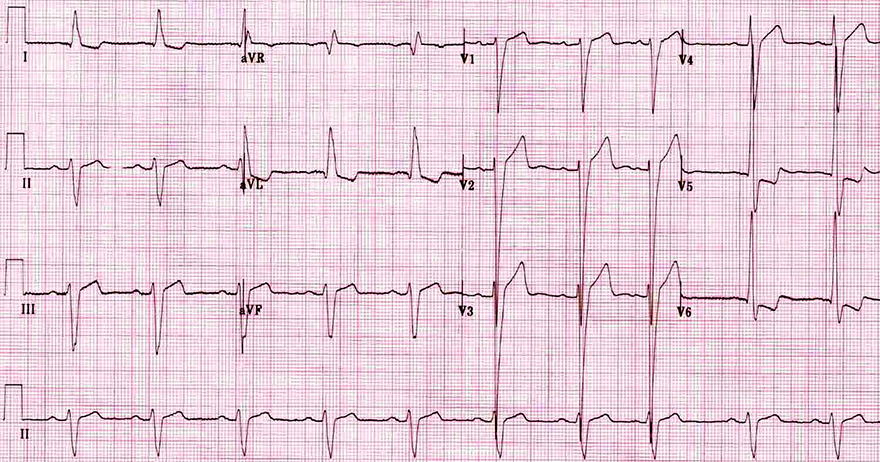
Left Ventricular Hypertrophy

  • Large S and R waves in precordial leads that extend into adjacent leads
  • Positive Sokolow Index
    • S(V1/2) + R(V5/6) ≥ 35mm
  • Left Axis Deviation
    • aVL(R) ≥ 13mm and III(S) ≥ 15mm
  • Left Ventricular Overload
    • Descending ST depressions in lateral leads (V5-V6, I, aVL)
    • Likely concentric hypertrophy of the LV
  • Discordant ST Elevations (V1-3)
  • Prolonged R Wave Peak Time (V6) ≥ 50ms
  • Definitive diagnosis is through echocardiography


ECG left ventricular hypertrophy, sokolov criteria, increased QRS voltages, discordance ST elevation, LV strain pattern

Left Ventricular Hypertrophy


Sources

  • ECG from Basics to Essentials Step by Step
  • litfl.com
  • ecgwaves.com
  • metealpaslan.com
  • medmastery.com
  • uptodate.com
  • ecgpedia.org
  • wikipedia.org
  • Strong Medicine
  • Understanding Pacemakers


šípka späť

Left Ventricular Hypertrophy

Left ventricular hypertrophy

Left Ventricle

  • The left ventricle has a physiological thickness of 10-12mm
  • Hemodynamic overload of the ventricle (volume or pressure) causes remodeling of the ventricle
    • Left ventricular hypertrophy
      • It is wall thickening > 12mm
      • The wall of the left ventricle has a physiological thickness of 10-12mm
    • Right ventricular hypertrophy
      • It is wall thickening > 5mm
      • The wall of the right ventricle has a physiological thickness of 3-4mm
  • According to hemodynamic overload, we recognize 2 types of hypertrophy:
    • Concentric hypertrophy
    • Excentric hypertrophy


Normal left ventricle thickness

Concentric Left Ventricular Hypertrophy

  • The ventricle is overloaded by pressure (increased afterload)
    • The ventricle pumps blood against a higher pressure
  • Hypertrophy without dilation of the ventricular cavity occurs
  • Concentric left ventricular hypertrophy arises with systolic overload of the left ventricle:
    • Arterial hypertension
    • Aortic stenosis
  • Concentric right ventricular hypertrophy occurs with systolic overload of the right ventricle:

left ventricle concentric hypertrophy

Eccentric Left Ventricular Hypertrophy

  • The ventricle is overloaded by volume (increased preload)
    • It becomes filled with blood during diastole
    • During systole, it is overloaded by pressure (pumps a larger volume of blood)
  • Dilation and hypertrophy of the ventricular cavity occurs
    • With chronic volume overload, only dilation occurs
  • Eccentric left ventricular hypertrophy, causes:
    • Aortic insufficiency
  • Eccentric right ventricular hypertrophy, causes:
    • Pulmonary insufficiency

left ventricle eccentric hypertrophy

Main Ventricular Vector and Ventricular Hypertrophy

  • The main ventricular vector is best "observed"
  • Ventricular hypertrophy is seen on the ECG
    • Best in leads (V1 and V6)

  • Left Ventricular Hypertrophy (LVH)
  • Right Ventricular Hypertrophy (RVH)
    • The cardiac vector rotates in the direction of RVH
    • The vector is smaller because the RV is smaller than the LV
    • The vector points almost directly
      • toward lead V1
      • away from lead V6
      • QRS amplitudes will be smaller
ECG pattern features V1, V6, left ventricular hypertrophy, right ventricular hypertrophy

Left Ventricular Hypertrophy
  • ECG cannot differentiate between concentric and eccentric hypertrophy
  • ECG in the diagnosis of left ventricular hypertrophy has:
    • Low sensitivity (20-50%)
      • Many patients with hypertrophy have a normal ECG (false negatives)
    • High specificity (90%)
      • If a patient has ECG signs of hypertrophy, then hypertrophy is very likely
  • Diagnosis of hypertrophy is performed using echocardiography
  • The principle of ECG diagnosis of LVH is that the cardiac vector rotates:
    • Toward lateral leads (I, aVL, V5-V6)
    • Away from right-sided leads (aVR, III, V1-V2)

ECG and Left Ventricular Hypertrophy


Sokolow Index

  • S(V1/2) + R(V5/6) ≥ 35mm
  • It is the most commonly used criterion

  • Cornell Index
    • More sensitive than the Sokolow index
    • Less commonly remembered (often not used)
    • Men: R(aVL) + S(V3) > 28mm
    • Women: R(aVL) + S(V3) > 20mm


ECG, voltage criteria, Sokolow Lyon criteria, index, left ventricular hypertrophy
  • Cornell Product Duration
    • Has the highest sensitivity
    • Less commonly remembered (often not used)
    • Cornell Index (mm) x QRS Duration (ms) > 2440 mm.ms

Lewis Index

  • I(R) + III(S) > 25mm

ECG, voltage criteria, Lewis index, left ventricular hypertrophy

aVL(R) ≥ 11mm


ECG, voltage criteria, left ventricular hypertrophy, R wave in aVL 11mm

aVL(R) ≥ 13mm and III(S) ≥ 15mm

ECG, voltage criteria, left ventricular hypertrophy, aVL(R) 13mm and III(S) 15mm

Prolonged R Wave Peak Time (V6) ≥ 50ms

  • The time from the start of the QRS complex to the peak of the R wave is referred to as:
    • R Wave Peak Time (Intrinsicoid Deflection)

  • Prolonged R Wave Peak Time (V6) ≥ 50ms
    • Is seen in left ventricular hypertrophy

  • In hypertrophy, the time for wall activation is prolonged
  • Thus, the distance between:

ECG R Wave Peak Time (Intrinsicoid Deflection), left ventricular hypertrophy


ECG left ventricular hypertrophy, voltage criteria, sokolov lyon ciretir, lewis index, r wave in aVL, increased r wave peak time

Left Ventricular Hypertrophy


Left Ventricular Overload and Concentric Hypertrophy of the LV

Left ventricular strain pattern

  • Only concentric hypertrophy of the LV (pressure overload) is created
    • Not eccentric hypertrophy of the LV (volume overload)

  • Descending ST depressions in lateral leads (I, aVL, V5-6)
ECG Left ventricular strain pattern, ST depression, T wave inversion in lateral leads


ECG left ventricular hypertrophy, voltage criteria, increased LV voltages, r wave peak time, LV strain pattern, ST elevation, left axis deviation

Left Ventricular Hypertrophy


ECG left ventricular hypertrophy (LVH), voltage criteria, increased LV voltages, r wave peak time, LV strain pattern, ST elevation, left axis deviation

Left Ventricular Hypertrophy

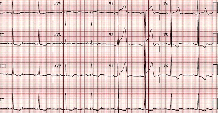
  • Large S and R waves in precordial leads that extend into adjacent leads
  • Positive Sokolow Index
    • S(V1/2) + R(V5/6) ≥ 35mm
  • Left Axis Deviation
    • aVL(R) ≥ 13mm and III(S) ≥ 15mm
  • Left Ventricular Overload
    • Descending ST depressions in lateral leads (V5-V6, I, aVL)
    • Likely concentric hypertrophy of the LV
  • Discordant ST Elevations (V1-3)
  • Prolonged R Wave Peak Time (V6) ≥ 50ms
  • Definitive diagnosis is through echocardiography


ECG left ventricular hypertrophy, sokolov criteria, increased QRS voltages, discordance ST elevation, LV strain pattern

Left Ventricular Hypertrophy


Sources

  • ECG from Basics to Essentials Step by Step
  • litfl.com
  • ecgwaves.com
  • metealpaslan.com
  • medmastery.com
  • uptodate.com
  • ecgpedia.org
  • wikipedia.org
  • Strong Medicine
  • Understanding Pacemakers