ECGbook.com

Making Medical Education Free for All

ECGbook.com

Making Medical Education Free for All

Home /

Memory T Waves

Cardiac memory, Memory induced T wave, Electrical remodeling, Chatterjee phenomenon (Post pacing T wave inversion)
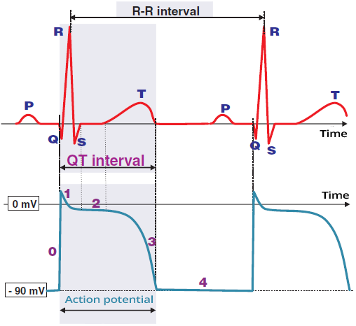
Action Potential and ECG

  • Action Potential (impulse) is a rapid change in the electrical voltage of cardiomyocytes
    • It arises from changes in the concentration of intracellular and extracellular ions
  • The action potential propagates

ECG action potential duration, T wave cardiac memory

ECG (Lead V6) and Action Potential

  • QRS Complex represents ventricular depolarization
    • During the formation of the QRS complex, a depolarization wave spreads through the ventricles
  • ST Segment is the isoelectric line
  • T Wave represents ventricular repolarization
    • The electrical vector during repolarization has the same direction as during depolarization
    • Repolarization, however, lasts longer, so the T wave is wider than the QRS complex
  • QT Interval corresponds to one cardiac cycle:
    • Depolarization + repolarization of the ventricles
  • P Wave represents atrial depolarization
    • The action potential of the P wave is not visible in the image
    • Atrial repolarization is hidden within the QRS complex

Cardiac Memory

Cardiac memory, Memory induced T wave, Electrical remodeling, Chatterjee phenomenon (Post pacing T wave inversion)

Cardiac Memory and ECG


cardiac memory, ventricular tachycardia ECG Memory induced T wave

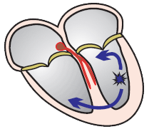
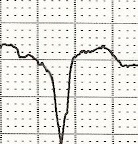
Wide QRS Complex

  • During abnormal ventricular depolarization
  • A cardiac memory may develop
    • Specifically in repolarization (T waves)


cardiac memory, sinus rhythm ECG Chatterjee phenomenon (Post pacing T wave inversion)

Sinus Rhythm and Memory T Waves

  • After a change from abnormal depolarization
  • The heart remembers the repolarization during the wide QRS
    • Memory inverted T waves develop

ECG and Memory T Waves


Heart cardiac memory, WPW syndrome, abnormal depolarization
ECG WPW syndrome, Cardiac memory, Memory induced T wave, Electrical remodeling

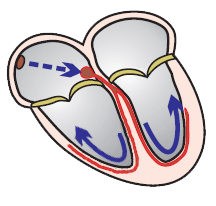
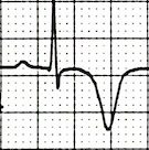
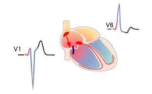
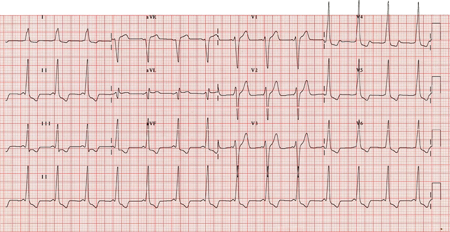
WPW Syndrome (Type B)

  • WPW Syndrome Type B
    • Shortened PQ interval < 0.12s
    • Delta wave and widened QRS approximately 0.15s
    • Negative QRS (V1)
      • This indicates that the Kent bundle is in the right atrium
  • Widened QRS complex
    • This represents abnormal ventricular depolarization
    • Secondarily, ventricular repolarization is also abnormal
      • (which the heart often remembers)


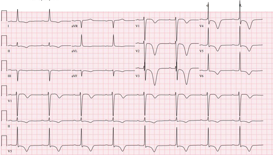
ECG cardiac memory T waves, after WPW syndrome accessory pathway ablation

Memory T Waves


ECG cardiac memory,  Memory induced T wave, Electrical remodeling, Chatterjee phenomenon (Post pacing T wave inversion)

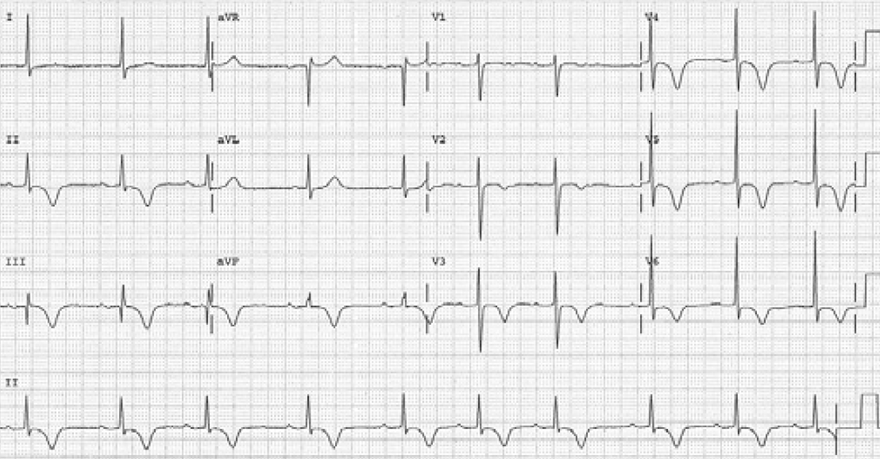
Memory T Waves

  • The patient has a pacemaker implanted for sick sinus syndrome
    • However, on this ECG, ventricular pacing is turned off
  • With sinus rhythm, memory T waves are present
    • Positive T wave (aVL)
    • Negative T waves (V1-V6)
    • Maximum negative T wave in precordial leads (V1-V6)
      • It is larger than the negative T wave (III)


Sources

  • ECG from Basics to Essentials Step by Step
  • litfl.com
  • ecgwaves.com
  • metealpaslan.com
  • medmastery.com
  • uptodate.com
  • ecgpedia.org
  • wikipedia.org
  • Strong Medicine
  • Understanding Pacemakers


šípka späť

Memory T Waves

Cardiac memory, Memory induced T wave, Electrical remodeling, Chatterjee phenomenon (Post pacing T wave inversion)

Action Potential and ECG

  • Action Potential (impulse) is a rapid change in the electrical voltage of cardiomyocytes
    • It arises from changes in the concentration of intracellular and extracellular ions
  • The action potential propagates

ECG action potential duration, T wave cardiac memory

ECG (Lead V6) and Action Potential

  • QRS Complex represents ventricular depolarization
    • During the formation of the QRS complex, a depolarization wave spreads through the ventricles
  • ST Segment is the isoelectric line
  • T Wave represents ventricular repolarization
    • The electrical vector during repolarization has the same direction as during depolarization
    • Repolarization, however, lasts longer, so the T wave is wider than the QRS complex
  • QT Interval corresponds to one cardiac cycle:
    • Depolarization + repolarization of the ventricles
  • P Wave represents atrial depolarization
    • The action potential of the P wave is not visible in the image
    • Atrial repolarization is hidden within the QRS complex

Cardiac Memory


Cardiac memory, Memory induced T wave, Electrical remodeling, Chatterjee phenomenon (Post pacing T wave inversion)

Cardiac Memory and ECG

cardiac memory, ventricular tachycardia br ECG Memory induced T wave

Wide QRS Complex

  • During abnormal ventricular depolarization
  • A cardiac memory may develop
    • Specifically in repolarization (T waves)
cardiac memory, sinus rhythm
ECG Chatterjee phenomenon (Post pacing T wave inversion)

Sinus Rhythm and Memory T Waves

  • After a change from abnormal depolarization
  • The heart remembers the repolarization during the wide QRS
    • Memory inverted T waves develop

ECG and Memory T Waves


ECG WPW syndrome, Cardiac memory, Memory induced T wave, Electrical remodeling

WPW Syndrome (Type B)

  • WPW Syndrome Type B
    • Shortened PQ interval < 0.12s
    • Delta wave and widened QRS approximately 0.15s
    • Negative QRS (V1)
      • This indicates that the Kent bundle is in the right atrium
  • Widened QRS complex
    • This represents abnormal ventricular depolarization
    • Secondarily, ventricular repolarization is also abnormal
      • (which the heart often remembers)
Heart cardiac memory, WPW syndrome, abnormal depolarization


ECG cardiac memory T waves, after WPW syndrome accessory pathway ablation

Memory T Waves


ECG cardiac memory,  Memory induced T wave, Electrical remodeling, Chatterjee phenomenon (Post pacing T wave inversion)

Memory T Waves

  • The patient has a pacemaker implanted for sick sinus syndrome
    • However, on this ECG, ventricular pacing is turned off
  • With sinus rhythm, memory T waves are present
    • Positive T wave (aVL)
    • Negative T waves (V1-V6)
    • Maximum negative T wave in precordial leads (V1-V6)
      • It is larger than the negative T wave (III)


Sources

  • ECG from Basics to Essentials Step by Step
  • litfl.com
  • ecgwaves.com
  • metealpaslan.com
  • medmastery.com
  • uptodate.com
  • ecgpedia.org
  • wikipedia.org
  • Strong Medicine
  • Understanding Pacemakers