ECGbook.com

Making Medical Education Free for All

ECGbook.com

Making Medical Education Free for All

Home /

Myocardial Infarction and aVR (Forgotten Lead)

Myocardial infarction and ST elevation in aVR (forgotten lead)
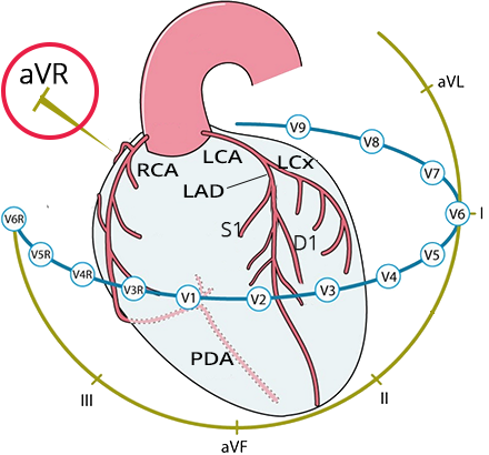
Lead aVR

Lead aVR and acute coronary syndrome
  • Lead aVR "looks at" the
    • Upper part of the right ventricle
    • Basal part of the ventricular septum
  • It has significant importance in diagnosing:
  • It is often overlooked in ACS
    • Hence it is referred to as the "forgotten lead"

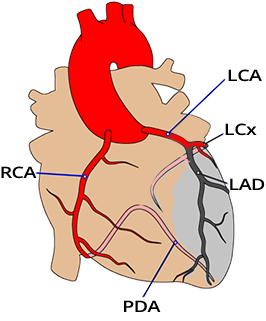
  • Basic coronary supply of the heart:
    • RCA (Right Coronary Artery)
    • LCA (Left Coronary Artery)
    • LAD (Left Anterior Descending)
    • LCx (Left Circumflex)
    • PDA (Posterior Descending Artery)

3 Major Arteries

Three major coronary heart arteries: Left Anterior Descending (LAD), Circumflex (LCx), Right Coronary Artery (RCA)

ST Elevation in aVR and Diffuse ST Depression

  • Acute Coronary Syndrome (ACS) with ST elevation in aVR and diffuse ST depressions
    • Has a very poor prognosis and the aVR lead is often overlooked

  • ST elevation in aVR has 4 causes:
    • Proximal Occlusion of the LAD
    • Occlusion of the LCA trunk
    • Stenosis of the LCA trunk
    • Triple Vessel Disease

Proximal Occlusion of the LAD

ST elevation in aVR, transmural infarction due to proximal LAD occlusion, basal septum infarction

Occlusion of the Left Main Coronary Artery

ST elevation in aVR, Left main coronary artery (LMCA) occlusion
  • Is very rare on the ECG, as patients almost always die suddenly
  • In the case of occlusion of the left main coronary artery
    • Causes an even larger infarct than in proximal LAD occlusion
    • Because both LAD and LCx are affected
  • Results in a massive infarct affecting:
    • Anterior wall (supplied by LAD) - ST elevations (V1-V4)
    • Lateral wall (supplied by both LAD and LCx) - ST elevations (I, aVL, V5-V6)
    • Posterior wall (partially supplied by LCx) - ST elevations (V7-V9)
    • Inferior wall (if LCx is dominant) - ST elevations (II, III, aVF)
  • The vectors of the individual walls affect each other
    • Resulting ischemic vector points away from the anterior heart
    • Causes ST depressions almost across the entire heart
  • Infarction of the ventricular septum due to occlusion of the first septal branch (S1)
    • Causes ST elevation in the aVR lead

  • ECG and occlusion of the left main coronary artery:
    • ST elevation in aVR ≥ 1mm
    • Diffuse ST depressions

Stenosis of the Left Main Coronary Artery

aVR ST elevation, Left main coronary artery (LMCA) insufficiency (stenosis)

Three-Vessel Disease

aVR ST elevation, Multi vessel artery disease, Triple vessel disease (3VD)
  • It is severe stenosis of all 3 major arteries
  • It leads to extensive subendocardial ischemia of the entire heart
  • Three-vessel disease and stenosis of the left main coronary artery have the same ECG pattern
    • Even though in three-vessel disease there is still stenosis of the right coronary artery
    • Small ischemic vector of the right ventricle
      • It is overlaid by a large vector of the massive left ventricle
  • The mechanism of ST elevation in aVR is exactly like in stenosis of the left main coronary artery

  • ECG and stenosis of the left main coronary artery:
    • ST elevation in aVR ≥ 1mm
    • Diffuse ST depressions

Coronary Artery Bypass Grafting (CABG)

NSTEMI with ST elevation in aVR, Coronary Artery Bypass Grafting

proximal LAD occlusion - culprit artery
ECG ST elevation in aVR, proximal LAD occlusion, widespread ST depression

Acute Proximal LAD Occlusion

  • The patient does not have the typical STEMI anterior wall presentation
  • ST elevation in aVR and V1
  • ST depressions (V3-V6, I, II, III, aVF)
  • The patient had an angiography showing proximal LAD occlusion

proximal LAD occlusion - culprit artery
ECG ST elevation in aVR, subacute septal STEMI, widespread ST depression

Acute Proximal LAD Occlusion


ST elevation in aVR, Left main coronary artery (LMCA) occlusion
ECG ST elevation (aVR), Left main coronary artery occlusion, ST elevation in aVR, widespread ST depression

Acute Occlusion of the Left Main Coronary Artery

  • ST elevation in aVR and V1
  • Diffuse ST depressions almost throughout the entire heart (V4-V6, I, II, aVL)
  • The patient had occlusion of the left main coronary artery on angiography
    • This is a rare ECG finding, as such a patient often dies suddenly
    • ECG is often not captured in time

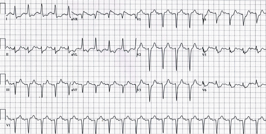
aVR ST elevation, Multi vessel artery disease, Triple vessel disease (3VD)
ECG diffuse subendocardial ischemia, severe triple vessel disease (SVD), ST elevation aVR

Triple Vessel Disease


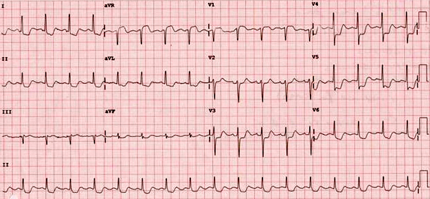
aVR ST elevation, Left main coronary artery (LMCA) insufficiency (stenosis)
ECG diffuse subendocardial ischemia, left main coronary artery stenosis

Stenosis of the Left Main Coronary Artery

  • ST elevation in aVR and V1
  • Diffuse ST depressions almost throughout the entire heart (V4-V6, I, II, aVL)
  • The patient had stenosis of the left main coronary artery on angiography

ST elevation in aVR, Left main coronary artery (LMCA) occlusion
ECG ST elevation (aVR), Left main coronary artery occlusion, ST elevation in aVR, widespread horizontal ST depression

Acute Occlusion of the Left Main Coronary Artery


ST elevation in aVR, Left main coronary artery (LMCA) occlusion
ECG ST elevation (aVR) occlusion left main stem, massive ST elevation - Pardee curve - wave

Acute Occlusion of the Left Main Coronary Artery

  • Massive ST elevations in almost all leads
  • Occlusion of the main artery can also cause a "classic STEMI picture"
  • The patient had an occlusion of the main dominant left coronary artery
    • This is a rare ECG finding because such a patient often dies suddenly
    • ECG is often not recorded in time


Sources

  • ECG from Basics to Essentials Step by Step
  • litfl.com
  • ecgwaves.com
  • metealpaslan.com
  • medmastery.com
  • uptodate.com
  • ecgpedia.org
  • wikipedia.org
  • Strong Medicine
  • Understanding Pacemakers


šípka späť

Myocardial Infarction and aVR (Forgotten Lead)

Myocardial infarction and ST elevation in aVR (forgotten lead)
Lead aVR

  • Lead aVR "looks at" the
    • Upper part of the right ventricle
    • Basal part of the ventricular septum
  • It has significant importance in diagnosing:
  • It is often overlooked in ACS
    • Hence it is referred to as the "forgotten lead"

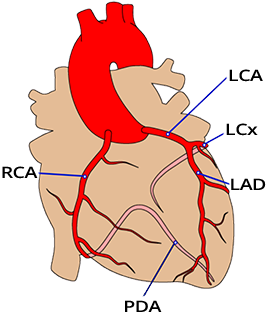
  • Basic coronary supply of the heart:
    • RCA (Right Coronary Artery)
    • LCA (Left Coronary Artery)
    • LAD (Left Anterior Descending)
    • LCx (Left Circumflex)
    • PDA (Posterior Descending Artery)

Lead aVR and acute coronary syndrome

3 Major Arteries

  • The heart is supplied by 3 major arteries:
    • Right Coronary Artery (RCA)
      • Posterior Descending Artery (PDA)
    • Left Coronary Artery (LCA):
      • Left Anterior Descending (LAD)
      • Left Circumflex (LCx)

  • PDA determines coronary artery dominance:
    • RCA Dominance (PDA branches from RCA)
    • LCA Dominance (PDA branches from LCA)
    • Codominance (PDA branches from both RCA and LCA)

  • Occlusion (blockage) of a major artery causes:
    • Infarction (necrosis) of the corresponding myocardium
  • Stenosis (narrowing) of a major artery causes:
    • Ischemia (up to subendocardial necrosis) of the corresponding myocardium

Three major coronary heart arteries: Left Anterior Descending (LAD), Circumflex (LCx), Right Coronary Artery (RCA)

ST Elevation in aVR and Diffuse ST Depression

  • Acute Coronary Syndrome (ACS) with ST elevation in aVR and diffuse ST depressions
    • Has a very poor prognosis and the aVR lead is often overlooked

  • ST elevation in aVR has 4 causes:
    • Proximal Occlusion of the LAD
    • Occlusion of the LCA trunk
    • Stenosis of the LCA trunk
    • Triple Vessel Disease

Proximal Occlusion of the LAD

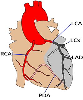
  • Proximal occlusion of the LAD above the S1 and D1 branches
  • Vectors of the anterior and lateral walls partially point away from each other
    • Thus they significantly change direction
    • Resulting ischemic vector points away from the anterior heart
    • This can cause ST depressions above the anterior and lateral walls
  • Infarction of the ventricular septum due to occlusion of the first septal branch (S1)
    • Causes ST elevation in the aVR lead

  • ECG and proximal occlusion of the LAD:
    • ST elevation in aVR ≥ 1mm
    • Diffuse ST depressions

ST elevation in aVR, transmural infarction due to proximal LAD occlusion, basal septum infarction

Occlusion of the Left Main Coronary Artery

  • Is very rare on the ECG, as patients almost always die suddenly
  • In the case of occlusion of the left main coronary artery
    • Causes an even larger infarct than in proximal LAD occlusion
    • Because both LAD and LCx are affected
  • Results in a massive infarct affecting:
    • Anterior wall (supplied by LAD) - ST elevations (V1-V4)
    • Lateral wall (supplied by both LAD and LCx) - ST elevations (I, aVL, V5-V6)
    • Posterior wall (partially supplied by LCx) - ST elevations (V7-V9)
    • Inferior wall (if LCx is dominant) - ST elevations (II, III, aVF)
  • The vectors of the individual walls affect each other
    • Resulting ischemic vector points away from the anterior heart
    • Causes ST depressions almost across the entire heart
  • Infarction of the ventricular septum due to occlusion of the first septal branch (S1)
    • Causes ST elevation in the aVR lead

  • ECG and occlusion of the left main coronary artery:
    • ST elevation in aVR ≥ 1mm
    • Diffuse ST depressions

ST elevation in aVR, Left main coronary artery (LMCA) occlusion

Stenosis of the Left Main Coronary Artery

  • In critical stenosis of the left main coronary artery, the following occurs:
  • Flow through the left main coronary artery is extremely low but maintained
    • Risk of occlusion of the left main coronary artery and sudden cardiac death
  • Mechanism of ST depressions across almost the entire heart is:
  • ST depressions (V4-V6, I, II, aVL) are anatomically opposite to the aVR lead
  • ECG and stenosis of the left main coronary artery:
    • ST elevation in aVR ≥ 1mm
    • Diffuse ST depressions

aVR ST elevation, Left main coronary artery (LMCA) insufficiency (stenosis)

Three-Vessel Disease

  • It is severe stenosis of all 3 major arteries
  • It leads to extensive subendocardial ischemia of the entire heart
  • Three-vessel disease and stenosis of the left main coronary artery have the same ECG pattern
    • Even though in three-vessel disease there is still stenosis of the right coronary artery
    • Small ischemic vector of the right ventricle
      • It is overlaid by a large vector of the massive left ventricle
  • The mechanism of ST elevation in aVR is exactly like in stenosis of the left main coronary artery

  • ECG and stenosis of the left main coronary artery:
    • ST elevation in aVR ≥ 1mm
    • Diffuse ST depressions

aVR ST elevation, Multi vessel artery disease, Triple vessel disease (3VD)

Coronary Artery Bypass Grafting (CABG)

  • Acute Coronary Syndrome with ST elevation in aVR
    • Has a very poor prognosis
    • Especially occlusion of the left main coronary artery
      • Without spontaneous recanalization, the patient suddenly dies

  • Patients with NSTEMI with ST elevation in aVR may have:
    • Stenosis of the left main coronary artery
    • Three-vessel disease

  • NSTEMI with aVR elevation
    • Requires cardiovascular surgical intervention
      • Placement of a coronary artery bypass graft
      • Therefore, these patients should not receive clopidogrel

NSTEMI with ST elevation in aVR, Coronary Artery Bypass Grafting


ECG ST elevation in aVR, proximal LAD occlusion, widespread ST depression

Acute Proximal LAD Occlusion
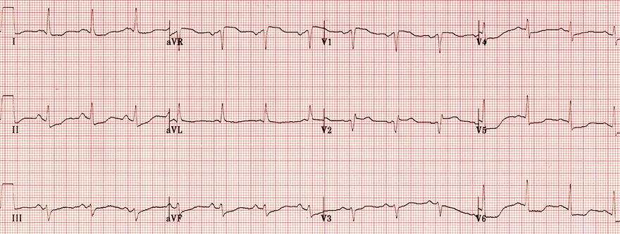
  • The patient does not have the typical STEMI anterior wall presentation
  • ST elevation in aVR and V1
  • ST depressions (V3-V6, I, II, III, aVF)
  • The patient had an angiography showing proximal LAD occlusion
proximal LAD occlusion - culprit artery

ECG ST elevation in aVR, subacute septal STEMI, widespread ST depression

Acute Proximal LAD Occlusion

  • The patient does not have the typical STEMI anterior wall presentation
  • ST elevation in aVR
  • ST depressions (I, II, III, aVF, V3-V6)
  • Subacute Septal STEMI
    • ST elevation and Q wave (V1-V2)
  • The patient had proximal LAD occlusion on angiography
proximal LAD occlusion - culprit artery

ECG ST elevation (aVR), Left main coronary artery occlusion, ST elevation in aVR, widespread ST depression

Acute Occlusion of the Left Main Coronary Artery

  • ST elevation in aVR and V1
  • Diffuse ST depressions almost throughout the entire heart (V4-V6, I, II, aVL)
  • The patient had occlusion of the left main coronary artery on angiography
    • This is a rare ECG finding, as such a patient often dies suddenly
    • ECG is often not captured in time
ST elevation in aVR, Left main coronary artery (LMCA) occlusion

ECG diffuse subendocardial ischemia, severe triple vessel disease (SVD), ST elevation aVR

Triple Vessel Disease

  • ST elevation in aVR
  • Diffuse ST depressions almost throughout the entire heart (V5-V6, I, II, aVL, aVF)
  • Subacute Septal STEMI
    • ST elevation and Q wave (V1-V2)
  • The patient had stenosis of 3 main vessels on angiography
aVR ST elevation, Multi vessel artery disease, Triple vessel disease (3VD)

ECG diffuse subendocardial ischemia, left main coronary artery stenosis

Stenosis of the Left Main Coronary Artery

  • ST elevation in aVR and V1
  • Diffuse ST depressions almost throughout the entire heart (V4-V6, I, II, aVL)
  • The patient had stenosis of the left main coronary artery on angiography
aVR ST elevation, Left main coronary artery (LMCA) insufficiency (stenosis)

ECG ST elevation (aVR), Left main coronary artery occlusion, ST elevation in aVR, widespread horizontal ST depression

Acute Occlusion of the Left Main Coronary Artery
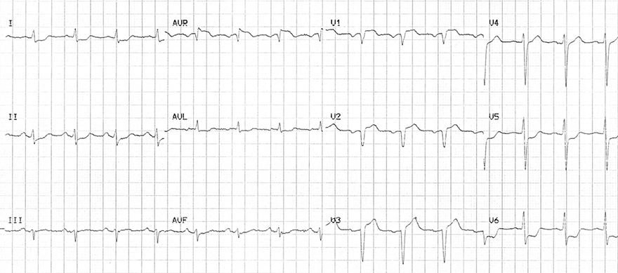
  • ST elevation in aVR and V1
  • Diffuse ST depressions almost across the entire heart (V3-V6, I, II, aVL)
  • The patient had occlusion of the left main coronary artery on angiography
    • This is a rare ECG finding because such a patient often dies suddenly
    • ECG is often not recorded in time
ST elevation in aVR, Left main coronary artery (LMCA) occlusion

ECG ST elevation (aVR) occlusion left main stem, massive ST elevation - Pardee curve - wave

Acute Occlusion of the Left Main Coronary Artery
  • Massive ST elevations in almost all leads
  • Occlusion of the main artery can also cause a "classic STEMI picture"
  • The patient had an occlusion of the main dominant left coronary artery
    • This is a rare ECG finding because such a patient often dies suddenly
    • ECG is often not recorded in time
ST elevation in aVR, Left main coronary artery (LMCA) occlusion


Sources

  • ECG from Basics to Essentials Step by Step
  • litfl.com
  • ecgwaves.com
  • metealpaslan.com
  • medmastery.com
  • uptodate.com
  • ecgpedia.org
  • wikipedia.org
  • Strong Medicine
  • Understanding Pacemakers