|
|
ECGbook.com Making Medical Education Free for All |
|
|
ECGbook.com Making Medical Education Free for All |
|
|
ECGbook.com Making Medical Education Free for All |


Sources
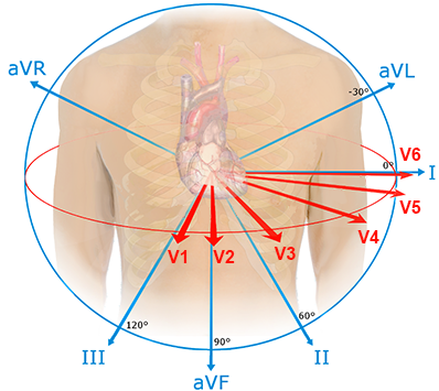
Standard 12-lead ECG
|
|
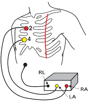
Modified Chest Lead
|
|
5-Electrode System
|
|
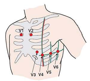
Leads V1 and V2 in the 6th Intercostal Space
|
|
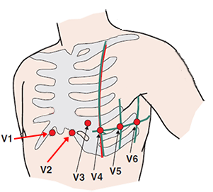
Leads V1 and V2 in the 2nd Intercostal Space
|
|
Lewis Lead
|
|
Esophageal ECG
|
|
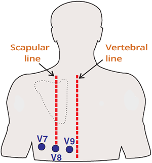
Posterior Leads (V7-V9)
|
|
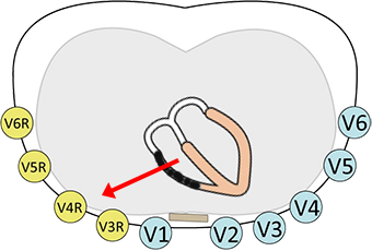
Right-Sided Leads (V4R-V6R)
|
|
Sources