|
|
ECGbook.com Making Medical Education Free for All |
Upload ECG for Interpretation |
|
|
ECGbook.com Making Medical Education Free for All |
Upload ECG for Interpretation |
|
|
ECGbook.com Making Medical Education Free for All |
Home /
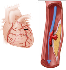
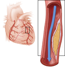
Non-ST Elevation Myocardial Infarction, NSTEMI Heart attack, Unstable angina pectoris
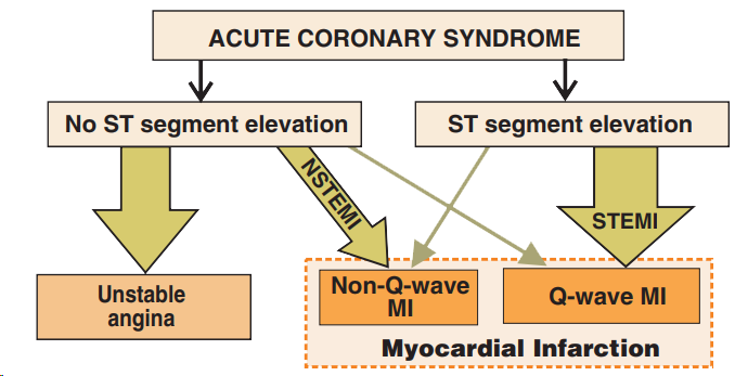
Acute Coronary Syndrome
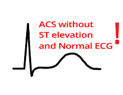
Acute Coronary Syndrome Without ST Elevation
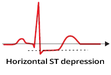

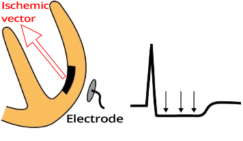
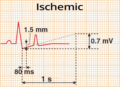
ST Depression and Ischemia
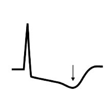
Flat Ascending ST Depression

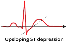
Steep Ascending ST Depression


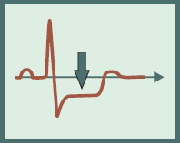
ST Depression

Negative T Waves

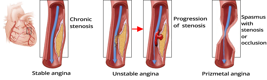
Unstable Angina Pectoris

Unstable Angina Pectoris

Ischemia Post-Ergometry
Unstable Angina Pectoris

Unstable Angina Pectoris

Pseudonormalization - Hyperacute STEMI
Sources
Non-ST Elevation Myocardial Infarction, NSTEMI Heart attack, Unstable angina pectoris
Acute Coronary Syndrome
|
|
|
|
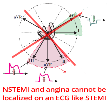
Acute Coronary Syndrome Without ST Elevation
|
|
|
|
ST Depression and Ischemia
|
Flat Ascending ST Depression
|
Steep Ascending ST Depression
|
Negative T Waves and ACS Without ST Elevation
|
|
|
|
|
|
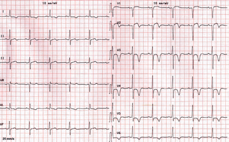
ST Depression
|
|
|
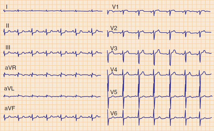
Negative T Waves
|
|
Unstable Angina Pectoris
|
|
|
Unstable Angina Pectoris
|
|
|
Ischemia Post-Ergometry
|
|
|
Unstable Angina Pectoris
|
|
|
Unstable Angina Pectoris
|
|
|
Pseudonormalization - Hyperacute STEMI
|
|
Sources