|
|
ECGbook.com Making Medical Education Free for All |
Upload ECG for Interpretation |
|
|
ECGbook.com Making Medical Education Free for All |
Upload ECG for Interpretation |
|
|
ECGbook.com Making Medical Education Free for All |
Home /
P cardiale, P biatriale, Biatrial enlargement (hypertrophy)
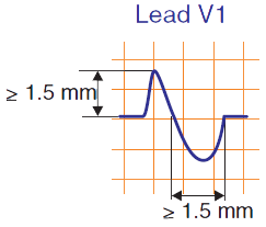
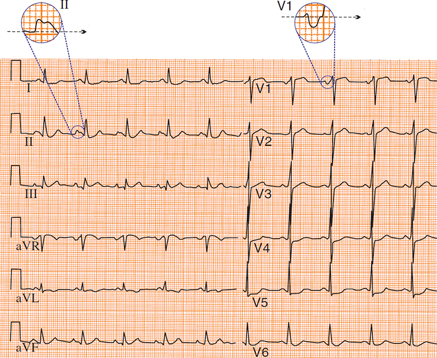
P Mitrale
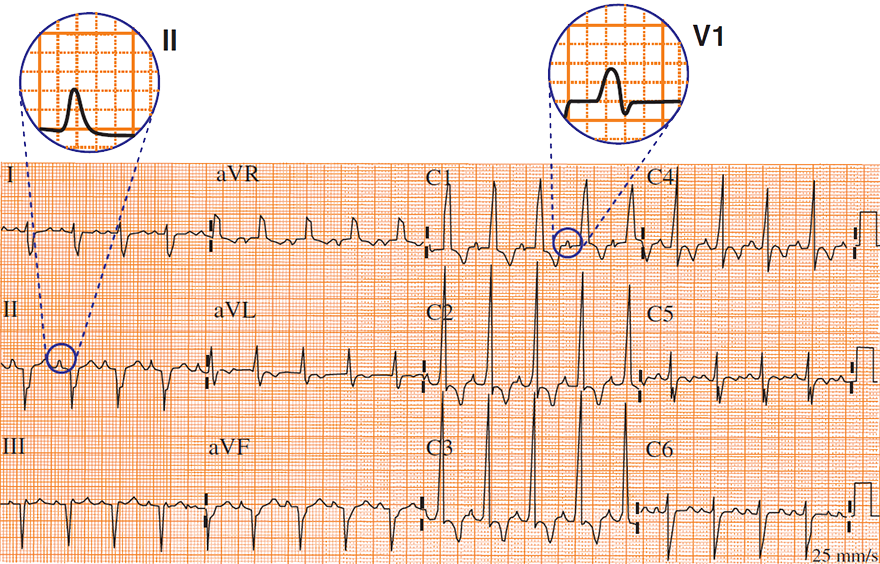
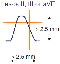
P Pulmonale
Biatrial Hypertrophy
Biatrial Hypertrophy
Sources
P cardiale, P biatriale, Biatrial enlargement (hypertrophy)
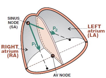
Physiological P Wave
|
|
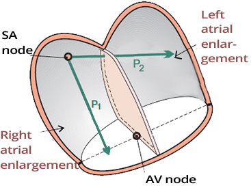
Biatrial Hypertrophy
|
|
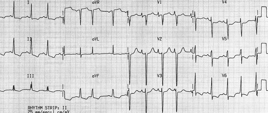
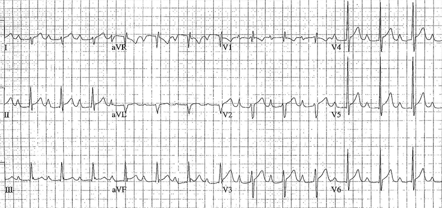
ECG and Biatrial Hypertrophy
|
|
P Mitrale
P Pulmonale
Biatrial Hypertrophy
Biatrial Hypertrophy
Sources