|
|
ECGbook.com Making Medical Education Free for All |
Upload ECG for Interpretation |
|
|
ECGbook.com Making Medical Education Free for All |
Upload ECG for Interpretation |
|
|
ECGbook.com Making Medical Education Free for All |
Home /
P dextrocardiale, Right atrial hypertrophy, Right atrial abnormality, Delay of right atrial activation, Right atrial dilatation, Right atrial distention, Right atrial overload
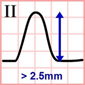
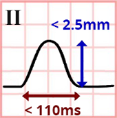
P Pulmonale and Lead II
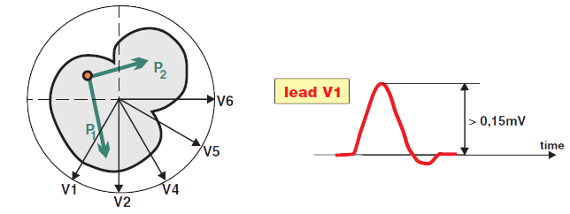
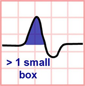
P Pulmonale and Lead V1

Normal P Wave
P Pulmonale
P Pulmonale
P Pulmonale

P Pulmonale

Cor Pulmonale (P Pulmonale + Right Ventricular Hypertrophy)
Sources
Home /
P dextrocardiale, Right atrial hypertrophy, Right atrial abnormality, Delay of right atrial activation, Right atrial dilatation, Right atrial distention, Right atrial overload
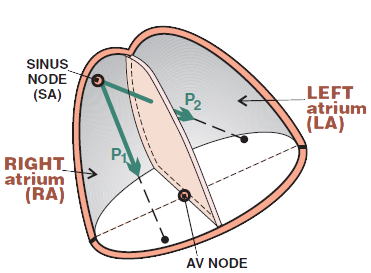
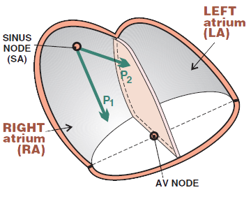
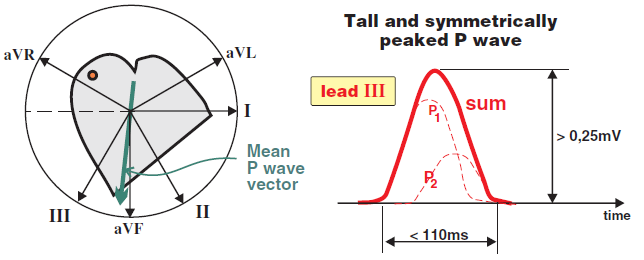
Physiological P Wave
|
|
|
|
|
|
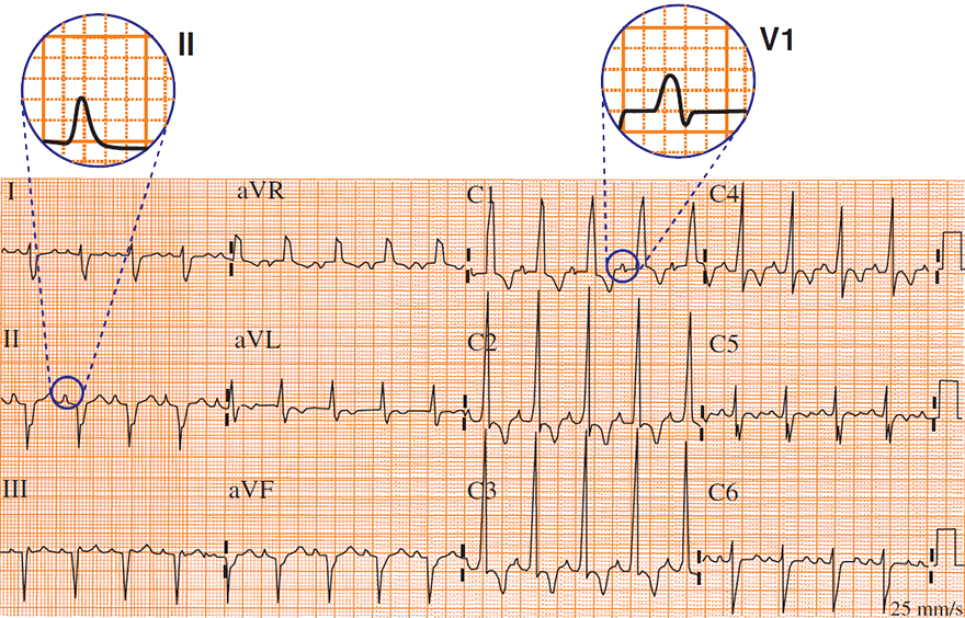
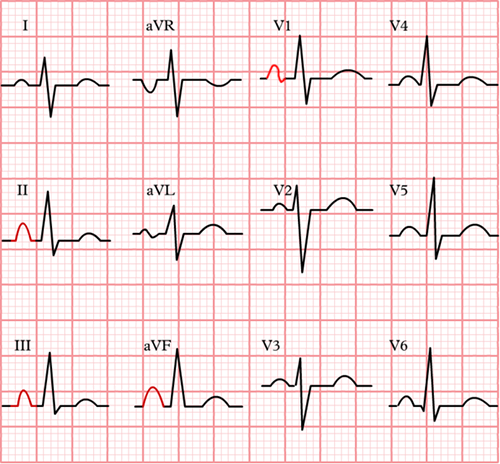
P Pulmonale and Lead II
P Pulmonale and Lead V1
ECG and P Pulmonale
|
|
|
|
|
|
Normal P Wave
|
|
|
|
P Pulmonale
P Pulmonale
P Pulmonale
P Pulmonale
Cor Pulmonale (P Pulmonale + Right Ventricular Hypertrophy)
Sources