|
|
ECGbook.com Making Medical Education Free for All |
|
|
ECGbook.com Making Medical Education Free for All |
|
|
ECGbook.com Making Medical Education Free for All |
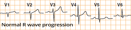
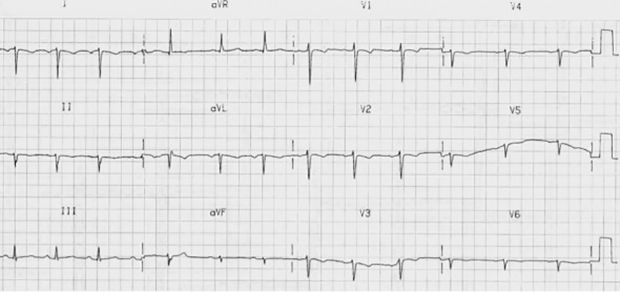
R Wave Progression and Transition Zone
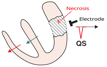
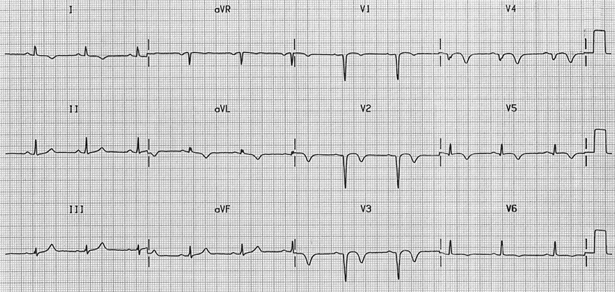
Amputated R Waves and Old Antero-septal Infarction
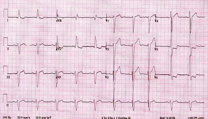
Decreased R Wave Progression and Left Ventricular Hypertrophy
Decreased R Wave Progression and Swapped Leads V1 and V3
Decreased R Wave Progression and Dilated Cardiomyopathy
Amputated R and Left Bundle Branch Block
WPW Syndrome Type B
Reduced R Wave Progression and Dextrocardia
Sources
|
|
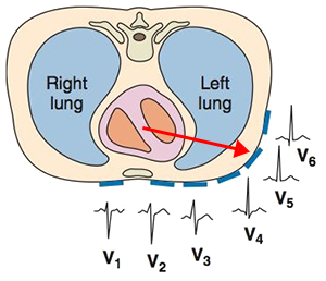
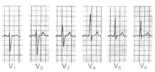
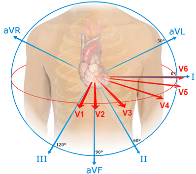
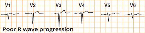
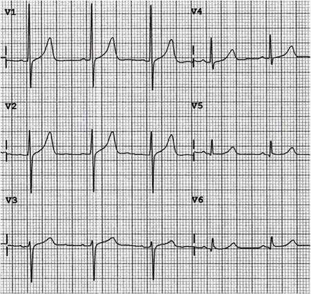
R Wave Progression and Transition Zone
Pathological R Wave
|
|
|
|
|
|
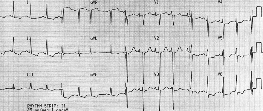
Amputated R Waves and Old Antero-septal Infarction
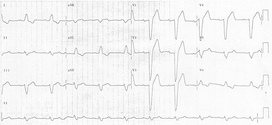
Decreased R Wave Progression and Left Ventricular Hypertrophy
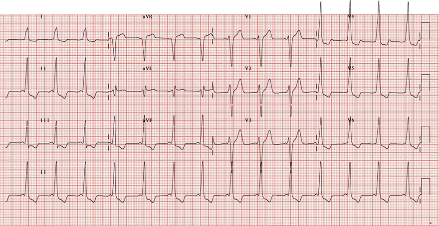
Decreased R Wave Progression and Swapped Leads V1 and V3
Decreased R Wave Progression and Dilated Cardiomyopathy
Amputated R and Left Bundle Branch Block
WPW Syndrome Type B
Reduced R Wave Progression and Dextrocardia
Sources