|
|
ECGbook.com Making Medical Education Free for All |
|
|
ECGbook.com Making Medical Education Free for All |
|
|
ECGbook.com Making Medical Education Free for All |


Acute Inferior, Posterior, and Lateral STEMI

Acute Inferior and Posterior STEMI


Acute Postero-Lateral STEMI

Old Posterior STEMI


Acute Inferior and Posterior STEMI

Acute STEMI?
Acute Posterior Wall STEMI

Subacute Inferior Posterior Lateral STEMI
Sources
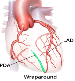
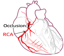
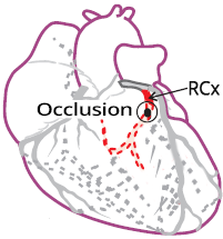
Coronary Artery Dominance
|
|
|
|
|
|
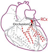
Posterior Wall Infarction
|
|
|
|
|
|
|
|
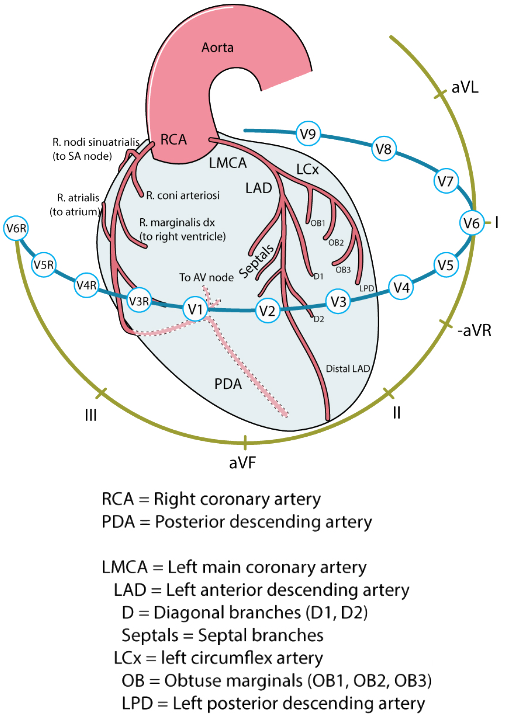
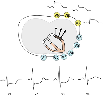
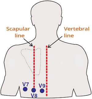
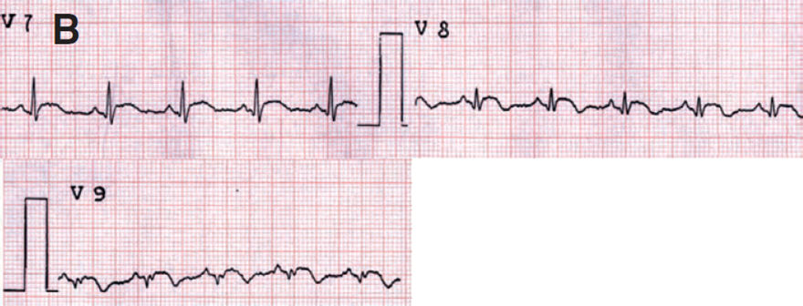
Posterior Leads (V7-V9)
|
|
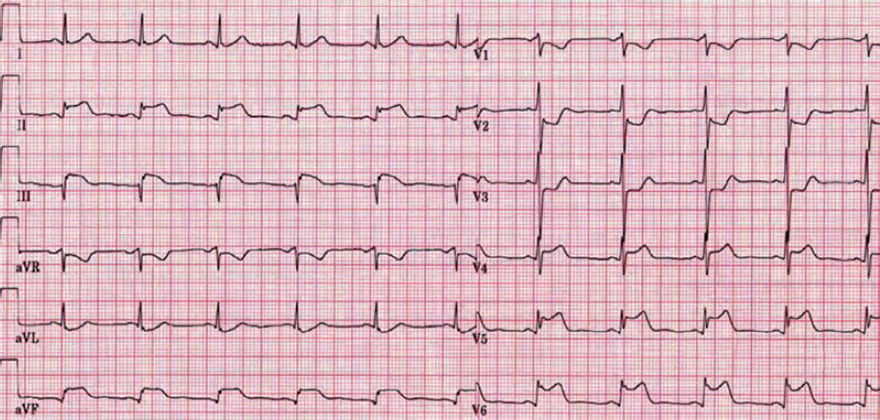
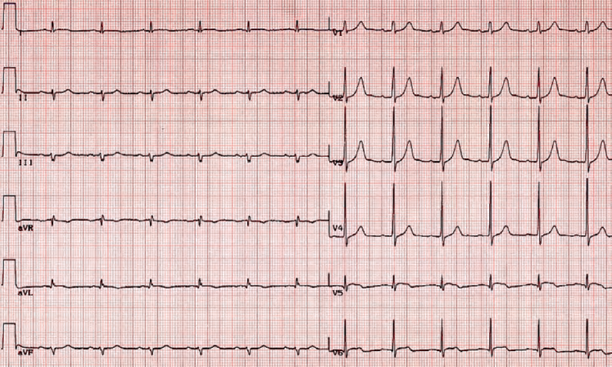
ECG and Posterior STEMI
|
|
|
Acute Inferior, Posterior, and Lateral STEMI
|
|
|
Acute Inferior and Posterior STEMI
|
|
|
Acute Postero-Lateral STEMI
|
|
|
Old Posterior STEMI
|
|
|
Acute Inferior and Posterior STEMI
|
|
|
Acute STEMI?
|
|
|
Acute Posterior Wall STEMI
|
|
|
Subacute Inferior Posterior Lateral STEMI
|
|
Sources