ECGbook.com

Making Medical Education Free for All

ECGbook.com

Making Medical Education Free for All

Home /

PQ Interval

PR (PQ) interval

PQ Interval (PR Interval)

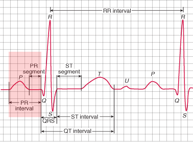
PR (PQ) interval from onset P wave to start of QRS complex
  • Interval is a section that contains waves or oscillations
  • Segment is a section that contains only the isoelectric line

  • PQ Interval
    • It is the section between the start of the P wave and the start of the QRS complex
    • The QRS complex most often starts with the Q wave
  • PR Interval
    • Sometimes the PQ interval is referred to as the PR interval
      • When the QRS complex starts with the R wave

  • Regardless of the configuration of the QRS complex
    • The PQ interval is more commonly used

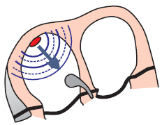
ECG Curve of the PQ Interval

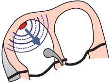
SA node atrial activation and PQ (PR) interval

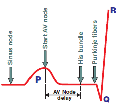
PQ (PR) interval and AV node conduction delay
  1. Impulse originates in the SA node
    • When it propagates to the atrial myocardium, the P wave begins to form
    • Simultaneously, it spreads through the conduction system toward the AV node
      • The impulse in the conduction system does not form a curve
  2. The impulse enters the AV node
    • The impulse spreads from the SA node
    • At the time of atrial activation (peak of the P wave)
      • It arrives via the conduction system to the AV node
  3. Delayed (decremental) conduction in the AV node
    • The impulse “lingers” in the AV node for about 0.1s (no curve is formed)
    • Then it passes to His bundle (no curve is formed)
  4. Activation of the ventricular septum
    • From His bundle the impulse through Purkinje fibers
      • Begins to activate the myocardium of the ventricular septum
      • Formation of the Q wave begins

ECG and PQ Interval

ECG normal PR (PQ) interval 0.12 - 0.2s

Shortened PQ Interval (<0.12s)


SA node, sinus tachycardia

ECG short PQ (PR) interval with sinus tachycardia

Sinus Tachycardia

  • Heart rate: 125/min
  • PQ Interval: 0.12s (4 squares)
    • The PQ interval is borderline shortened
  • In tachycardia, the PQ interval shortens
    • Conduction through the AV node speeds up


AV node depolarization (activation) and accelerated junctional rhythm

ECG accelerated junctional rhythm, inverted (retrograde) P waves with short PQ (PR) interval

Junctional Rhythm


WPW syndrome, accessory pathway - Bundle of Kent and short PQ (PR)

ECG WPW syndrome. Short PQ (PR) interval, broad QRS, delta wave

WPW Syndrome

  • PQ Interval 0.12s (3 squares)
  • In WPW syndrome, the ventricles are simultaneously activated via
  • Ventricles are activated earlier (pre-excited) via the Bundle of Kent
    • A delta wave forms, which shortens the PQ interval


Lown Ganong Levine syndrome with short PQ (PR) interval

ECG Lown Ganong Levine (LGL) syndrome. Short PQ (PR) interval, normal P wave, normal QRS complex, absent delta wave

LGL Syndrome


Prolonged PQ Interval (>0.2s)


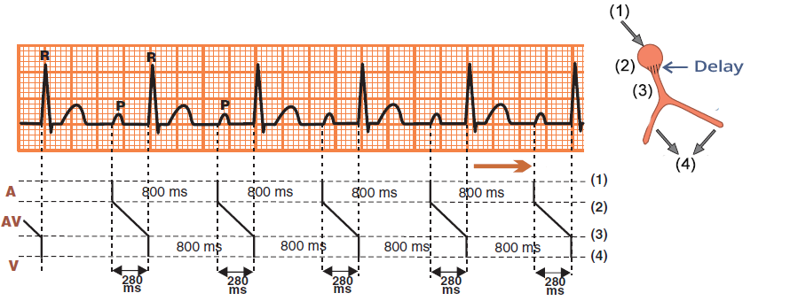
ECG and ladder diagram (laddergram) with first degree av block. Prolonged PQ (PR) interval, delayed conduction through the av node

First-Degree AV Block


Variable PQ Interval


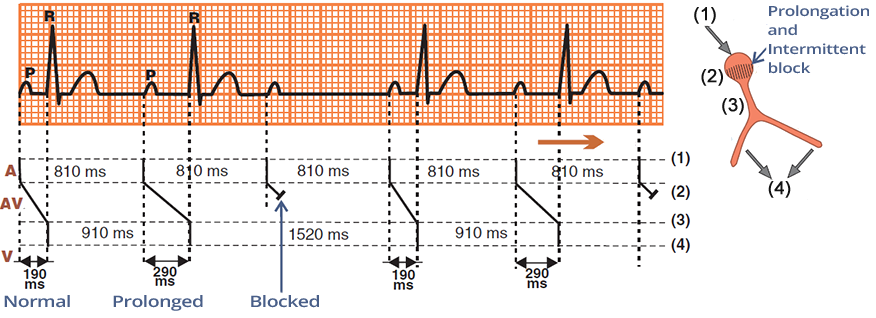
ECG (Ladder diagram - Laddergarm) with second degree AV block (Mobitz I - Wenckebach) with prolonged variable PQ (PR) interval

Second-Degree AV Block - Mobitz I (Wenckebach)
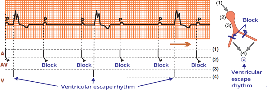


ECG (Ladder diagram - Laddergarm) 3rd degree AV block (complete heart block) variable PQ (PR interval), AV dissociation

Third-Degree AV Block and Ventricular Rhythm

  • Laddergram illustrates the propagation of the impulse through the conduction system
    • A - Atria (1), AV - AV Junction (2), V - Ventricles (4)
  • The SA node generates impulses regularly
  • A secondary pacemaker has activated in the ventricles, resulting in ventricular rhythm
    • QRS frequency: 35/min.
  • This is Third-Degree AV Block (Complete Block)
    • AV dissociation - P waves are independent of QRS complexes
    • PQ interval changes


Sources

  • ECG from Basics to Essentials Step by Step
  • litfl.com
  • ecgwaves.com
  • metealpaslan.com
  • medmastery.com
  • uptodate.com
  • ecgpedia.org
  • wikipedia.org
  • Strong Medicine
  • Understanding Pacemakers


šípka späť

PQ Interval

PR (PQ) interval

PQ Interval (PR Interval)

  • Interval is a section that contains waves or oscillations
  • Segment is a section that contains only the isoelectric line

  • PQ Interval
    • It is the section between the start of the P wave and the start of the QRS complex
    • The QRS complex most often starts with the Q wave
  • PR Interval
    • Sometimes the PQ interval is referred to as the PR interval
      • When the QRS complex starts with the R wave

  • Regardless of the configuration of the QRS complex
    • The PQ interval is more commonly used

PR (PQ) interval from onset P wave to start of QRS complex

ECG Curve of the PQ Interval

  1. Impulse originates in the SA node
    • When it propagates to the atrial myocardium, the P wave begins to form
    • Simultaneously, it spreads through the conduction system toward the AV node
      • The impulse in the conduction system does not form a curve
  2. The impulse enters the AV node
    • The impulse spreads from the SA node
    • At the time of atrial activation (peak of the P wave)
      • It arrives via the conduction system to the AV node
  3. Delayed (decremental) conduction in the AV node
    • The impulse “lingers” in the AV node for about 0.1s (no curve is formed)
    • Then it passes to His bundle (no curve is formed)
  4. Activation of the ventricular septum
    • From His bundle the impulse through Purkinje fibers
      • Begins to activate the myocardium of the ventricular septum
      • Formation of the Q wave begins
SA node atrial activation and PQ (PR) interval


PQ (PR) interval and AV node conduction delay

ECG and PQ Interval


ECG normal PR (PQ) interval 0.12 - 0.2s

Shortened PQ Interval (<0.12s)


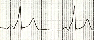
SA node, sinus tachycardia ECG short PQ (PR) interval with sinus tachycardia

Sinus Tachycardia

  • Heart rate: 125/min
  • PQ Interval: 0.12s (4 squares)
    • The PQ interval is borderline shortened
  • In tachycardia, the PQ interval shortens
    • Conduction through the AV node speeds up


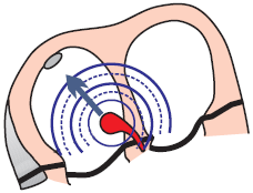
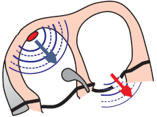
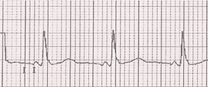
AV node depolarization (activation) and accelerated junctional rhythm ECG accelerated junctional rhythm, inverted (retrograde) P waves with short PQ (PR) interval

Junctional Rhythm

  • Heart rate: 50/min
  • PQ Interval: 0.08s (2 squares)
  • In junctional rhythm, the impulse originates in the AV junction
    • Atria and ventricles are activated almost simultaneously
      • Therefore, there is a shortened PQ interval
    • The atria are activated retrogradely (from bottom to top), resulting in:


WPW syndrome, accessory pathway - Bundle of Kent and short PQ (PR) ECG WPW syndrome. Short PQ (PR) interval, broad QRS, delta wave

WPW Syndrome

  • PQ Interval 0.12s (3 squares)
  • In WPW syndrome, the ventricles are simultaneously activated via
  • Ventricles are activated earlier (pre-excited) via the Bundle of Kent
    • A delta wave forms, which shortens the PQ interval


Lown Ganong Levine syndrome with short PQ (PR) interval ECG Lown Ganong Levine (LGL) syndrome. Short PQ (PR) interval, normal P wave, normal QRS complex, absent delta wave

LGL Syndrome

  • PQ Interval 0.08s (2 squares)
  • LGL Syndrome features a connection between the atria and the AV junction
    • The connection (James bundle) bypasses the AV node
    • The impulse does not slow down in the AV node
    • Therefore, the PQ interval will be shortened

Prolonged PQ Interval (>0.2s)


ECG and ladder diagram (laddergram) with first degree av block. Prolonged PQ (PR) interval, delayed conduction through the av node

First-Degree AV Block


Variable PQ Interval


ECG (Ladder diagram - Laddergarm) with second degree AV block (Mobitz I - Wenckebach) with prolonged variable PQ (PR) interval

Second-Degree AV Block - Mobitz I (Wenckebach)


ECG (Ladder diagram - Laddergarm) 3rd degree AV block (complete heart block) variable PQ (PR interval), AV dissociation

Third-Degree AV Block and Ventricular Rhythm

  • Laddergram illustrates the propagation of the impulse through the conduction system
    • A - Atria (1), AV - AV Junction (2), V - Ventricles (4)
  • The SA node generates impulses regularly
  • A secondary pacemaker has activated in the ventricles, resulting in ventricular rhythm
    • QRS frequency: 35/min.
  • This is Third-Degree AV Block (Complete Block)
    • AV dissociation - P waves are independent of QRS complexes
    • PQ interval changes


Sources

  • ECG from Basics to Essentials Step by Step
  • litfl.com
  • ecgwaves.com
  • metealpaslan.com
  • medmastery.com
  • uptodate.com
  • ecgpedia.org
  • wikipedia.org
  • Strong Medicine
  • Understanding Pacemakers