|
|
ECGbook.com Making Medical Education Free for All |
|
|
ECGbook.com Making Medical Education Free for All |
|
|
ECGbook.com Making Medical Education Free for All |


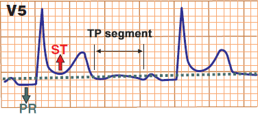
ST Elevations and PQ Depressions
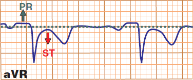
ST Depressions and PQ Elevations

Acute Pericarditis
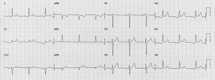
Acute Pericarditis
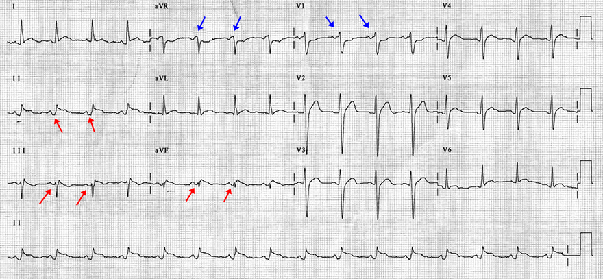
Atrial Infarction and Inferior Wall STEMI
Sources
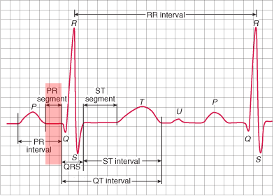
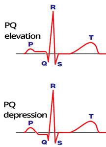
PQ Segment (PR Segment)
|
|
ECG Curve of the PQ Interval
|
|
ECG and PQ Segment
|
|
|
ST Elevations and PQ Depressions
|
ST Depressions and PQ Elevations
|
Acute Pericarditis
Acute Pericarditis
Atrial Infarction
|
|
Atrial Infarction and Inferior Wall STEMI
Sources