|
|
ECGbook.com Making Medical Education Free for All |
Upload ECG for Interpretation |
|
|
ECGbook.com Making Medical Education Free for All |
Upload ECG for Interpretation |
|
|
ECGbook.com Making Medical Education Free for All |
Home /
Premature ventricular complex (PVC), Ventricular premature beat (VPB), Ventricular extrasystole (VES), Premature ventricular contraction, Ventricular premature depolarisation, Ventricular ectopic
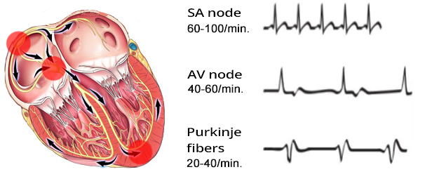
Basic Heart Rhythms
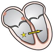
Discordance and Ventricular Extrasystole
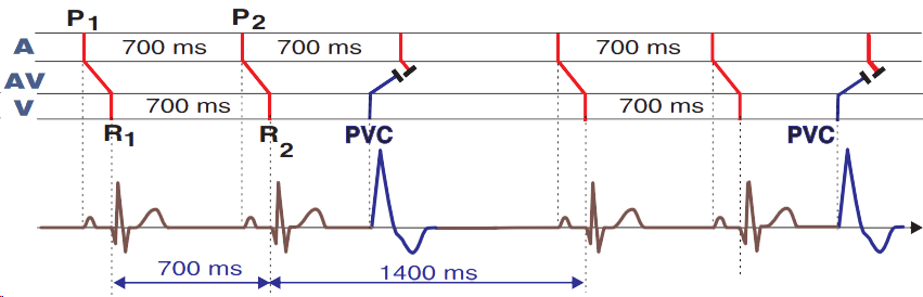
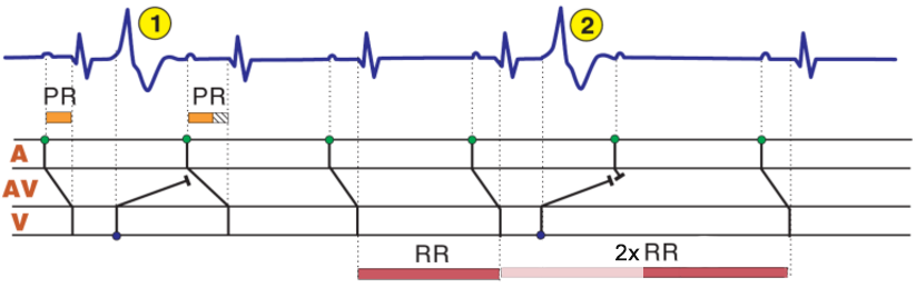
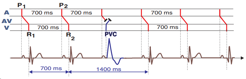
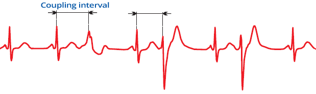
Full Compensatory Pause and Ventricular Extrasystole
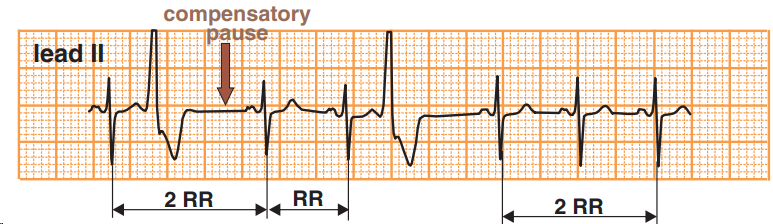
Full Compensatory Pause and Ventricular Extrasystole
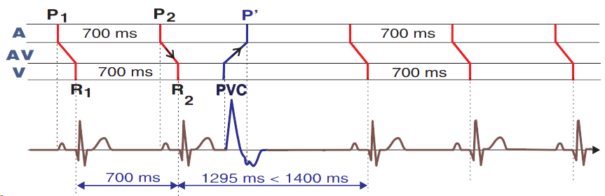
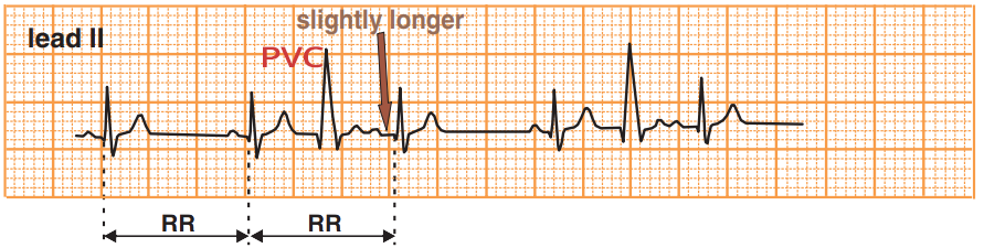
Incomplete Compensatory Pause and Ventricular Extrasystole
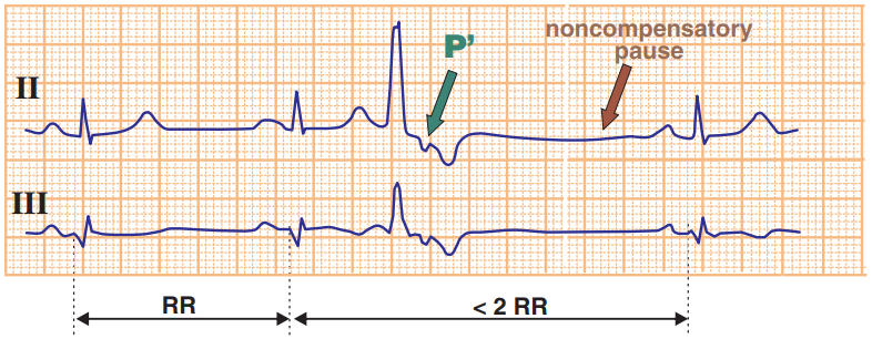
Incomplete Compensatory Pause and Ventricular Extrasystole
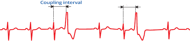
Interpolated Ventricular Extrasystole
Interpolated VES
End-Diastolic PVC
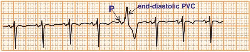
End-Diastolic PVC
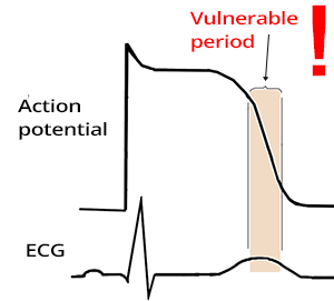
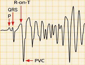
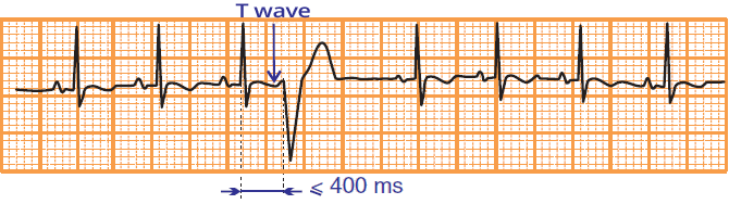
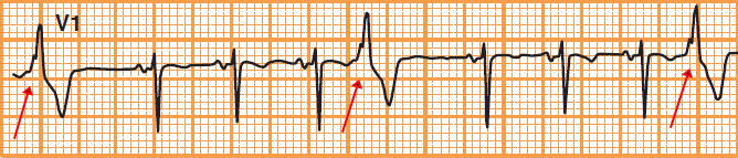
R-on-T Phenomenon (PVC on T Wave)
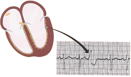
Focus is in the Right Ventricle at the Base of the Heart
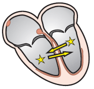
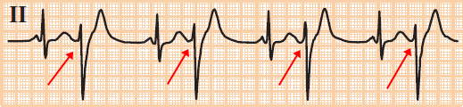
Unifocal PVC
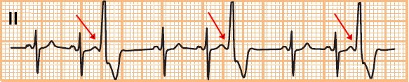
Multifocal PVC
Bigeminy
Trigeminy
Quadrigeminy
Couplet
Triplet (Salvo)
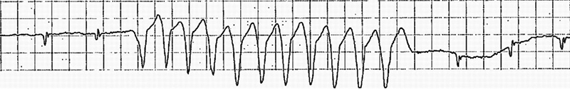
Non-Sustained Ventricular Tachycardia
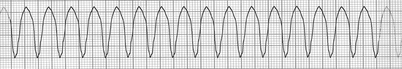
Sustained Ventricular Tachycardia
| Grade | Type of PVC |
| 0 | No PVCs |
| I | Unifocal PVCs <30/min |
| II | Unifocal PVCs ≥ 30/min |
| III | Multifocal PVCs |
| IVA | Couplets |
| IVB | Triplets Non-sustained ventricular tachycardia |
| V | R-on-T Phenomenon |
Sources
Home /
Premature ventricular complex (PVC), Ventricular premature beat (VPB), Ventricular extrasystole (VES), Premature ventricular contraction, Ventricular premature depolarisation, Ventricular ectopic
Basic Heart Rhythms
|
|
Discordance and Ventricular Extrasystole
Full Compensatory Pause and Ventricular Extrasystole
Full Compensatory Pause and Ventricular Extrasystole
Incomplete Compensatory Pause and Ventricular Extrasystole
Incomplete Compensatory Pause and Ventricular Extrasystole
|
|
Interpolated Ventricular Extrasystole
Interpolated VES
End-Diastolic PVC
End-Diastolic PVC
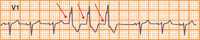
R-on-T Phenomenon
|
|
R-on-T Phenomenon (PVC on T Wave)
|
|
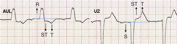
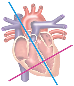
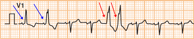
1. Chest Leads (V1-6): Left / Right Ventricle?
2. Inferior Leads (II, III, aVF): Apex / Base of the Heart?
|
|
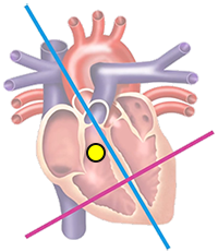
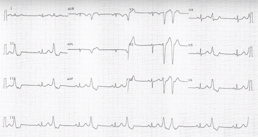
Focus is in the Right Ventricle at the Base of the Heart
|
|
|
|
|
Unifocal PVC
|
|
|
Multifocal PVC
Bigeminy
Trigeminy
Quadrigeminy
Couplet
Triplet (Salvo)
Non-Sustained Ventricular Tachycardia
Sustained Ventricular Tachycardia
| Grade | Type of PVC |
| 0 | No PVCs |
| I | Unifocal PVCs <30/min |
| II | Unifocal PVCs ≥ 30/min |
| III | Multifocal PVCs |
| IVA | Couplets |
| IVB | Triplets Non-sustained ventricular tachycardia |
| V | R-on-T Phenomenon |
Sources